
-
Product Name
MDH1 antibody
- Documents
-
Description
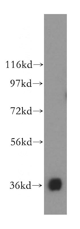
MDH1 Rabbit Polyclonal antibody. Positive IP detected in mouse liver tissue. Positive WB detected in HL-60 cells, HepG2 cells, human kidney tissue, human spleen tissue, mouse liver tissue. Positive FC detected in HepG2 cells. Positive IF detected in HepG2 cells. Positive IHC detected in human renal cell carcinoma tissue, human liver cancer tissue. Observed molecular weight by Western-blot: 36 kDa
-
Tested applications
ELISA, WB, IF, FC, IP, IHC
-
Species reactivity
Human,Mouse,Rat; other species not tested.
-
Alternative names
Cytosolic malate dehydrogenase antibody; MDH s antibody; MDH1 antibody; MDHA antibody; MOR2 antibody
-
Isotype
Rabbit IgG
-
Preparation
This antibody was obtained by immunization of MDH1 recombinant protein (Accession Number: NM_005917). Purification method: Antigen affinity purified.
-
Clonality
Polyclonal
-
Formulation
PBS with 0.02% sodium azide and 50% glycerol pH 7.3.
-
Storage instructions
Store at -20℃. DO NOT ALIQUOT
-
Applications
Recommended Dilution:
WB: 1:500-1:5000
IP: 1:500-1:5000
IHC: 1:50-1:500
IF: 1:10-1:100
-
Validations
HL-60 cells were subjected to SDS PAGE followed by western blot with Catalog No:112568(MDH1 antibody) at dilution of 1:500
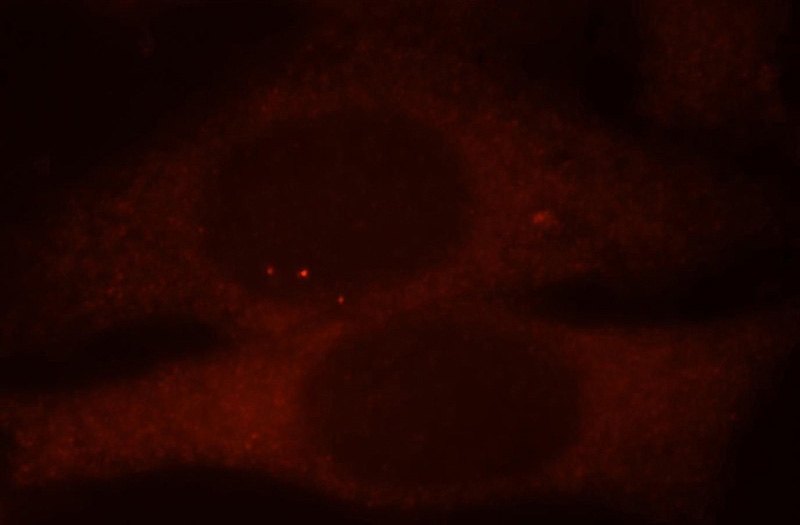
Immunofluorescent analysis of HepG2 cells, using MDH1 antibody Catalog No:112568 at 1:25 dilution and Rhodamine-labeled goat anti-rabbit IgG (red).
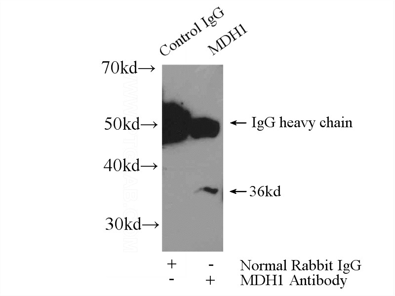
IP Result of anti-MDH1 (IP:Catalog No:112568, 4ug; Detection:Catalog No:112568 1:1000) with mouse liver tissue lysate 6400ug.
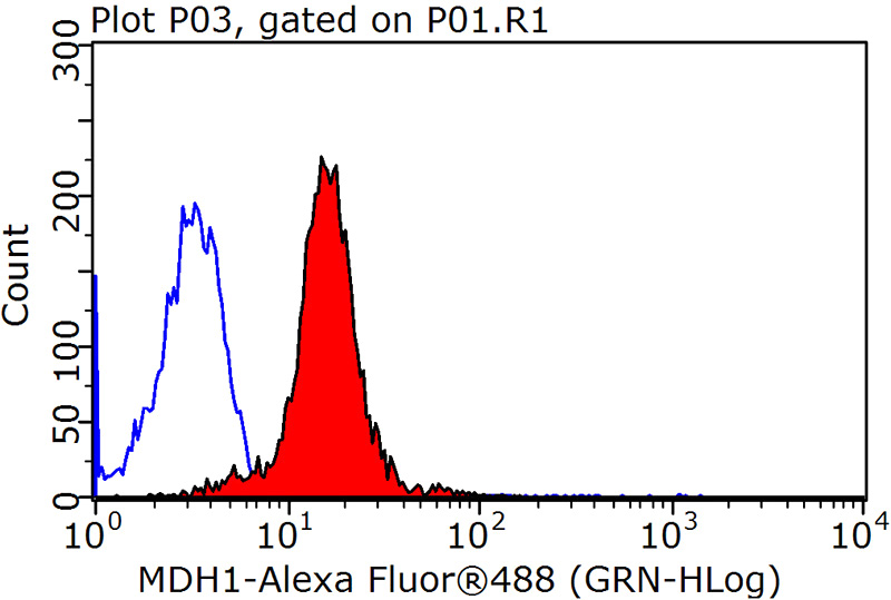
1X10^6 HepG2 cells were stained with 0.2ug MDH1 antibody (Catalog No:112568, red) and control antibody (blue). Fixed with 90% MeOH blocked with 3% BSA (30 min). Alexa Fluor 488-congugated AffiniPure Goat Anti-Rabbit IgG(H+L) with dilution 1:1500.
-
Background
MDH1(Malate dehydrogenase, cytoplasmic) is also named as MDHA and belongs to the LDH/MDH superfamily and MDH type 2 family which catalyzes the reversible oxidation of malate to oxaloacetate, utilizing the NAD/NADH cofactor system in the citric acid cycle. It can exsit as a dimer and the dimeric MDH1 is the mitochondrial isoenzyme, whereas the tetrameric MDH2 is the glycosomal isoenzyme.(PMID:10693743).
-
References
- Birsoy K, Wang T, Chen WW, Freinkman E, Abu-Remaileh M, Sabatini DM. An Essential Role of the Mitochondrial Electron Transport Chain in Cell Proliferation Is to Enable Aspartate Synthesis. Cell. 162(3):540-51. 2015.
- Dai J, Xu W, Zhao X. Protein profile screening: reduced expression of Sord in the mouse epididymis induced by nicotine inhibits tyrosine phosphorylation level in capacitated spermatozoa. Reproduction (Cambridge, England). 151(3):227-37. 2016.
- Ma X, Li C, Sun L. Lin28/let-7 axis regulates aerobic glycolysis and cancer progression via PDK1. Nature communications. 5:5212. 2014.
- Sun L, Song L, Wan Q. cMyc-mediated activation of serine biosynthesis pathway is critical for cancer progression under nutrient deprivation conditions. Cell research. 25(4):429-44. 2015.
Related Products / Services
Please note: All products are "FOR RESEARCH USE ONLY AND ARE NOT INTENDED FOR DIAGNOSTIC OR THERAPEUTIC USE"