
-
Product Name
GFAP antibody
- Documents
-
Description
GFAP Rabbit Polyclonal antibody. Positive WB detected in C6 cells, mouse brain tissue. Positive IHC detected in human brain tissue, human cerebellum tissue, human gliomas tissue, mouse brain tissue, rat brain tissue, rat cerebellum tissue. Observed molecular weight by Western-blot: 50 kDa
-
Tested applications
ELISA, WB, IHC
-
Species reactivity
Human,Mouse,Rat; other species not tested.
-
Alternative names
FLJ45472 antibody; GFAP antibody
-
Isotype
Rabbit IgG
-
Preparation
This antibody was obtained by immunization of GFAP recombinant protein (Accession Number: NM_002055). Purification method: Antigen affinity purified.
-
Clonality
Polyclonal
-
Formulation
PBS with 0.02% sodium azide and 50% glycerol pH 7.3.
-
Storage instructions
Store at -20℃. DO NOT ALIQUOT
-
Applications
Recommended Dilution:
WB: 1:500-1:5000
IHC: 1:500-1:4000
-
Validations
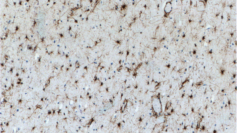
Immunohistochemistry of paraffin-embedded human brain tissue slide using Catalog No:110945(GFAP Antibody) at dilution of 1:2000 (under 10x lens).
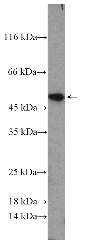
C6 cells were subjected to SDS PAGE followed by western blot with Catalog No:110945(GFAP Antibody) at dilution of 1:1000
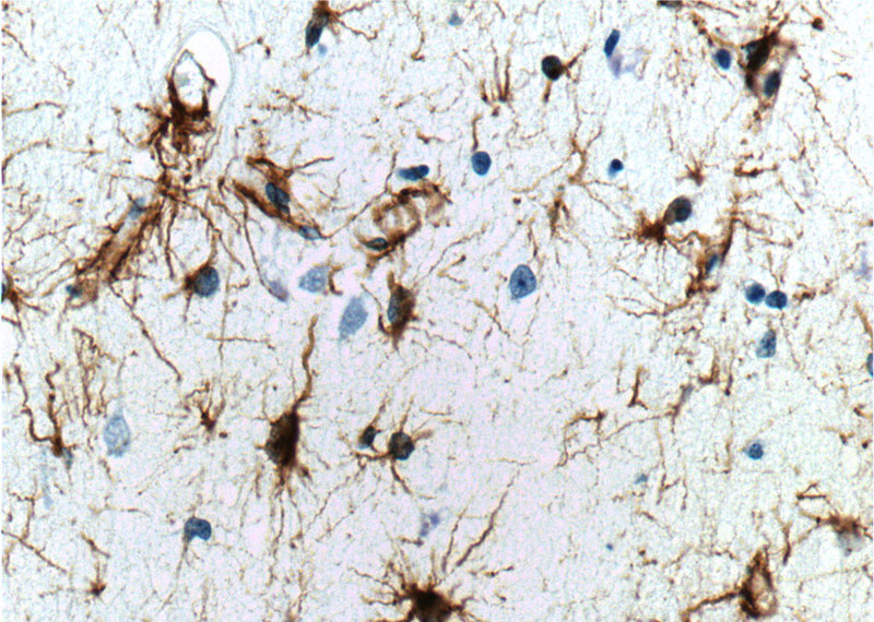
Immunohistochemistry of paraffin-embedded human brain tissue slide using Catalog No:110945(GFAP Antibody) at dilution of 1:2000 (under 40x lens).
-
Background
GFAP (Glial fibrillary acidic protein ), an intermediate-filament (IF) protein , is specifically expressed in cells of astroglial lineage and is widely used to mark astroglia in the brain. It is also used as a marker for intracranial and intraspinal tumors arising from astrocytes.
-
References
- Mohlin C, Liljekvist-Soltic I, Johansson K. Further assessment of neuropathology in retinal explants and neuroprotection by human neural progenitor cells. Journal of neural engineering. 8(6):066012. 2011.
- Zhang P, Men J, Fu Y. Contribution of SATB2 to the stronger osteogenic potential of bone marrow stromal cells from craniofacial bones. Cell and tissue research. 350(3):425-37. 2012.
- Zhang SY, Li BY, Li XL. Effects of phlorizin on diabetic retinopathy according to isobaric tags for relative and absolute quantification-based proteomics in db/db mice. Molecular vision. 19:812-21. 2013.
- Wang N, Zhang Y, Wu L. Puerarin protected the brain from cerebral ischemia injury via astrocyte apoptosis inhibition. Neuropharmacology. 79:282-9. 2014.
- Liu X, Yang Z, Yin Y, Deng X. Increased expression of Notch1 in temporal lobe epilepsy: animal models and clinical evidence. Neural regeneration research. 9(5):526-33. 2014.
- Anderson SR, Lee I, Ebeling C. Disrupted SOX10 function causes spongiform neurodegeneration in gray tremor mice. Mammalian genome : official journal of the International Mammalian Genome Society. 26(1-2):80-93. 2015.
- Zhu J, Mu X, Zeng J. Ginsenoside Rg1 prevents cognitive impairment and hippocampus senescence in a rat model of D-galactose-induced aging. PloS one. 9(6):e101291. 2014.
- Genové G, Mollick T, Johansson K. Photoreceptor degeneration, structural remodeling and glial activation: a morphological study on a genetic mouse model for pericyte deficiency. Neuroscience. 279:269-84. 2014.
Related Products / Services
Please note: All products are "FOR RESEARCH USE ONLY AND ARE NOT INTENDED FOR DIAGNOSTIC OR THERAPEUTIC USE"