
-
Product Name
Anti-Xrcc2 antibody
- Documents
-
Description
Rabbit polyclonal antibody to Xrcc2
-
Tested applications
WB, IHC-P
-
Species reactivity
Mouse, Human
-
Alternative names
Re antibody; RecA antibody; RAD51 antibody; 4921524O04Rik antibody; 8030409M04Rik antibody
-
Isotype
Rabbit IgG
-
Preparation
This antigen of this antibody was recombinant protein within human xrcc2 aa 1-200.
-
Clonality
Polyclonal
-
Formulation
Liquid, 1*TBS (pH7.4), 0.2% BSA, 50% Glycerol. Preservative: 0.05% Sodium Azide.
-
Storage instructions
Store at +4℃ after thawing. Aliquot store at -20℃. Avoid repeated freeze / thaw cycles.
-
Applications
WB: 1:500-1:2,000
IHC-P: 1:100-1:500
-
Validations
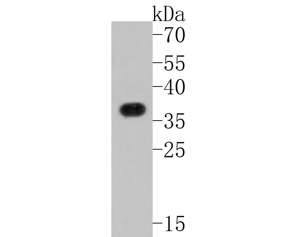
Fig1:; Western blot analysis of XRCC2 on mouse brain tissue lysates. Proteins were transferred to a PVDF membrane and blocked with 5% BSA in PBS for 1 hour at room temperature. The primary antibody ( 1/500) was used in 5% BSA at room temperature for 2 hours. Goat Anti-Rabbit IgG - HRP Secondary Antibody (HA1001) at 1:5,000 dilution was used for 1 hour at room temperature.
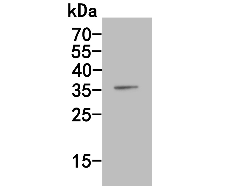
Fig2:; Western blot analysis of XRCC2 on human kidney tissue lysates. Proteins were transferred to a PVDF membrane and blocked with 5% BSA in PBS for 1 hour at room temperature. The primary antibody ( 1/500) was used in 5% BSA at room temperature for 2 hours. Goat Anti-Rabbit IgG - HRP Secondary Antibody (HA1001) at 1:5,000 dilution was used for 1 hour at room temperature.
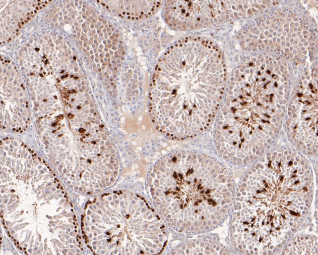
Fig3:; Immunohistochemical analysis of paraffin-embedded mouse testis tissue using anti-XRCC2 antibody. The section was pre-treated using heat mediated antigen retrieval with sodium citrate buffer (pH 6.0) for 20 minutes. The tissues were blocked in 5% BSA for 30 minutes at room temperature, washed with ddH; 2; O and PBS, and then probed with the primary antibody ( 1/400) for 30 minutes at room temperature. The detection was performed using an HRP conjugated compact polymer system. DAB was used as the chromogen. Tissues were counterstained with hematoxylin and mounted with DPX.
- Background
-
References
- Kluźniak W. et. al. Inherited variants in XRCC2 and the risk of breast cancer. Breast Cancer Res Treat. 2019 Dec
- Andreassen PR. et. al. XRCC2 (X-ray repair cross complementing 2). Atlas Genet Cytogenet Oncol Haematol. 2019 Jan
Related Products / Services
Please note: All products are "FOR RESEARCH USE ONLY AND ARE NOT INTENDED FOR DIAGNOSTIC OR THERAPEUTIC USE"