
-
Product Name
Anti-Phospho-Histone H2A.X (Ser139) (2A9) Mouse antibody
- Documents
-
Description
Phospho-Histone H2A.X (Ser139) (2A9) Mouse monoclonal antibody
-
Tested applications
WB, ICC/IF
-
Species reactivity
Human, Mouse
-
Alternative names
H2A histone family antibody; member X;H2A.X;H2a/x;H2AFX;H2AX;H2AX histone;H2AX_HUMAN;Histone H2A.X;Histone H2AX antibody
-
Isotype
Mouse IgG2a
-
Preparation
Antigen: Synthetic phosphopeptide corresponding to residues surrounding Ser139 of human H2A.X.
-
Clonality
Monoclonal
-
Formulation
Purified mouse monoclonal antibody in PBS(pH 7.4) containing with 0.03% Proclin300 and 50% glycerol.
-
Storage instructions
Store at 4°C short term. Store at -20°C long term. Avoid freeze / thaw cycle.
-
Applications
WB: 1/2000
;ICC: 1/400;Other 1/400
-
Validations
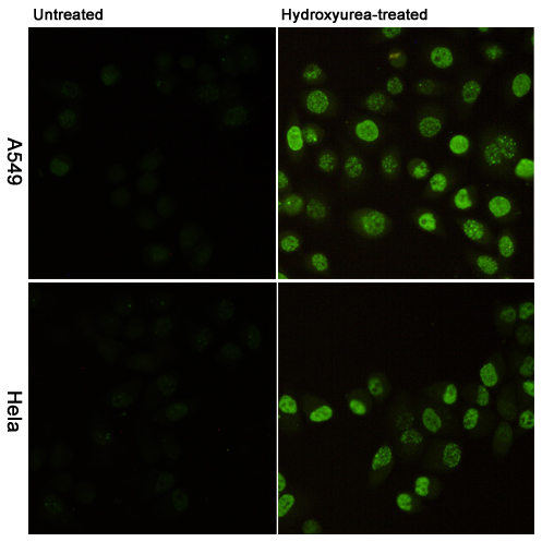
Immunofluorescent analysis of Phosphorylation of H2A.X at Serine 139 in A549(upper,untreated or Hydroxyurea-treated) and Hela(lower,untreated or Hydroxyurea-treated) using Phospho-Histone H2A.X (Ser139) mouse mAb (1:400).
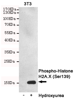
Western blot detection of Phosphorylation of H2A.X at Serine 139 in 3T3 or Hydroxyurea-treated 3T3 cell lysates using Phospho-Histone H2A.X (Ser139) mouse mAb (1:2000 diluted).Predicted band size:15KDa.Observed band size:15KDa.
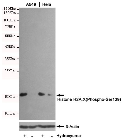
Western blot analysis of extracts from untreated or Hydroxyurea-treated Hela and A549 cells, using Histone H2A.X(Phospho-Ser139) mouse mAb (1:1000 diluted) (upper) or u03b2-Actin Mouse mAb (200068-8F10) (lower).Predicted band size:15KDa.Observed band size:15KDa.
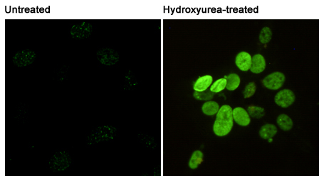
Immunofluorescent analysis of Phosphorylation of H2A.X at Serine 139 in 3T3 or Hydroxyurea-treated 3T3 cells using Phospho-Histone H2A.X
-
Background
Swiss-Prot Acc.P16104.Variant histone H2A which replaces conventional H2A in a subset of nucleosomes. Nucleosomes wrap and compact DNA into chromatin, limiting DNA accessibility to the cellular machineries which require DNA as a template. Histones thereby play a central role in transcription regulation, DNA repair, DNA replication and chromosomal stability. DNA accessibility is regulated via a complex set of post-translational modifications of histones, also called histone code, and nucleosome remodeling. Required for checkpoint-mediated arrest of cell cycle progression in response to low doses of ionizing radiation and for efficient repair of DNA double strand breaks (DSBs) specifically when modified by C-terminal phosphorylation.
Related Products / Services
Please note: All products are "FOR RESEARCH USE ONLY AND ARE NOT INTENDED FOR DIAGNOSTIC OR THERAPEUTIC USE"