
-
Product Name
Anti-H2BC12 antibody
- Documents
-
Description
Rabbit monoclonal antibody to H2BC12
-
Tested applications
WB, ICC/IF, IHC-P, IP, FC
-
Species reactivity
Human, Mouse, Rat
-
Alternative names
H2BK antibody; H2B/S antibody; H2BFT antibody; H2BFAiii antibody; HIST1H2BK antibody
-
Isotype
Rabbit IgG
-
Preparation
This antigen of this antibody was recombinant protein
-
Clonality
Monoclonal
-
Formulation
Liquid, 1*TBS (pH7.4), 0.05% BSA, 40% Glycerol. Preservative: 0.05% Sodium Azide.
-
Storage instructions
Store at +4℃ after thawing. Aliquot store at -20℃ or -80℃. Avoid repeated freeze / thaw cycles.
-
Applications
WB: 1:1,000
ICC/IF: 1:100-1:500
IHC-P: 1:100-1:500
FC: 1:50-1:100
-
Validations
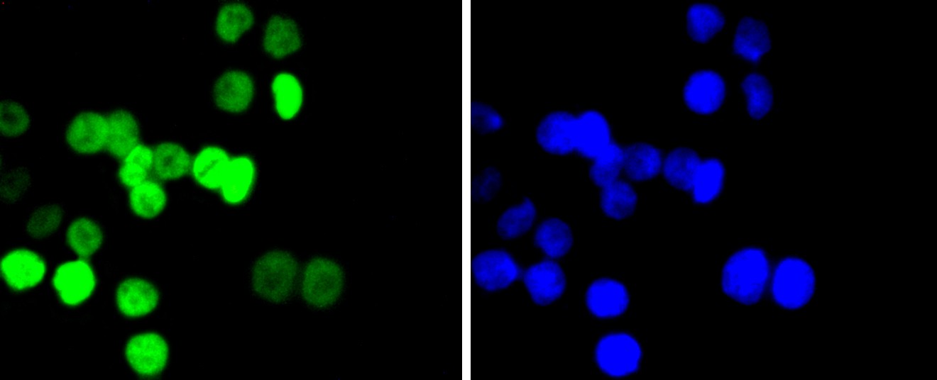
Fig1: ICC staining Histone H2B(acetyl K20) in SW480 cells (green). The nuclear counter stain is DAPI (blue). Cells were fixed in paraformaldehyde, permeabilised with 0.25% Triton X100/PBS.
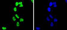
Fig2: ICC staining Histone H2B(acetyl K20) in Hela cells (green). The nuclear counter stain is DAPI (blue). Cells were fixed in paraformaldehyde, permeabilised with 0.25% Triton X100/PBS.
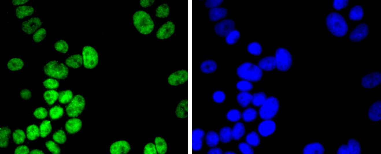
Fig3: ICC staining Histone H2B(acetyl K20) in MCF-7 cells (green). The nuclear counter stain is DAPI (blue). Cells were fixed in paraformaldehyde, permeabilised with 0.25% Triton X100/PBS.
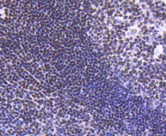
Fig4: Immunohistochemical analysis of paraffin-embedded human tonsil tissue using anti-Histone H2B(acetyl K20) antibody. Counter stained with hematoxylin.
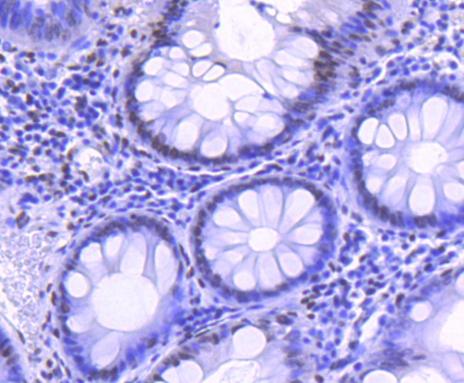
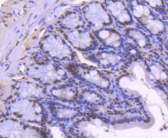
Fig5: Immunohistochemical analysis of paraffin-embedded human colon tissue using anti-Histone H2B(acetyl K20) antibody. Counter stained with hematoxylin.
Fig6: Immunohistochemical analysis of paraffin-embedded human colon cancer tissue using anti-Histone H2B(acetyl K20) antibody. Counter stained with hematoxylin.
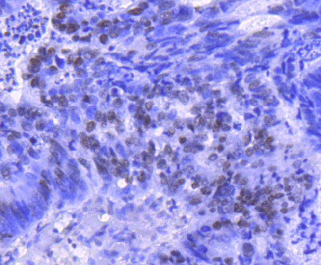
Fig7: Immunohistochemical analysis of paraffin-embedded human breast carcinoma tissue using anti-Histone H2B(acetyl K20) antibody. Counter stained with hematoxylin.
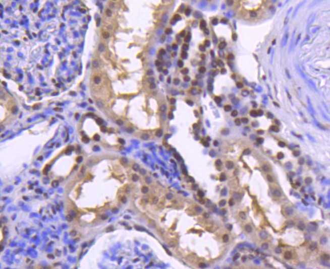
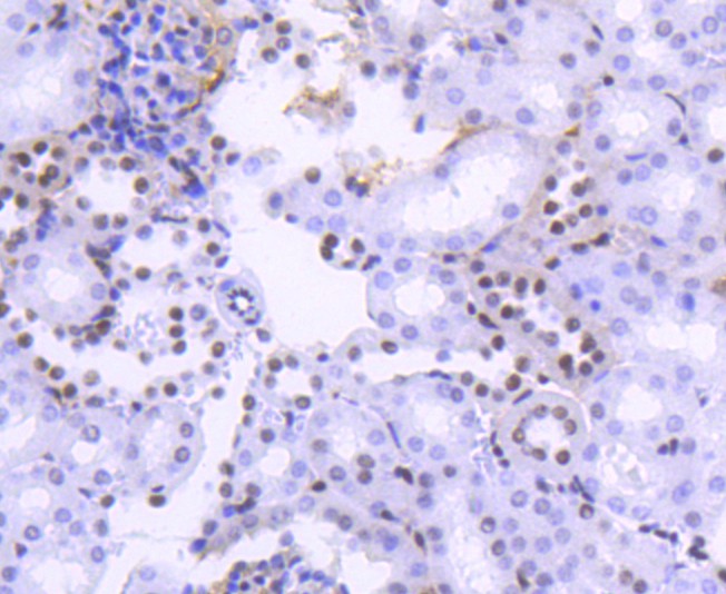
Fig8: Immunohistochemical analysis of paraffin-embedded human kidney tissue using anti-Histone H2B(acetyl K20) antibody. Counter stained with hematoxylin.
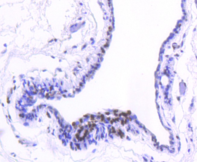
Fig9: Immunohistochemical analysis of paraffin-embedded mouse colon tissue using anti-Histone H2B(acetyl K20) antibody. Counter stained with hematoxylin.
Fig10: Immunohistochemical analysis of paraffin-embedded mouse kidney tissue using anti-Histone H2B(acetyl K20) antibody. Counter stained with hematoxylin.
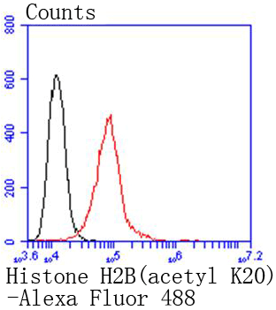
Fig11: Flow cytometric analysis of Hela cells with Histone H2B(acetyl K20) antibody at 1/50 dilution (red) compared with an unlabelled control (cells without incubation with primary antibody; black). Alexa Fluor 488-conjugated goat anti rabbit IgG was use
- Background
-
References
- Zhang B et al. An integrated peptide-antigen microarray on plasmonic gold films for sensitive human antibody profiling. PLoS One 8:e71043 (2013).
- Price JV et al. On silico peptide microarrays for high-resolution mapping of antibody epitopes and diverse protein-protein interactions. Nat Med : (2012).
Related Products / Services
Please note: All products are "FOR RESEARCH USE ONLY AND ARE NOT INTENDED FOR DIAGNOSTIC OR THERAPEUTIC USE"