
-
Product Name
Anti-Cyclin A2 Mouse antibody
- Documents
-
Description
Cyclin A2 Mouse monoclonal antibody
-
Tested applications
WB, IHC-P, FC
-
Species reactivity
Human
-
Isotype
Mouse IgG
-
Preparation
Antigen: Purified recombinant fragment of human CCNA2 (AA: 105-233) expressed in E. Coli.
-
Clonality
Monoclonal
-
Formulation
Purified antibody in PBS with 0.05% sodium azide.
-
Storage instructions
Store at 4°C short term. Store at -20°C long term. Avoid freeze / thaw cycle.
-
Applications
WB: 1/500 - 1/2000
IHC: 1/200 - 1/1000
FC: 1/200 - 1/400
ELISA: 1/10000
-
Validations
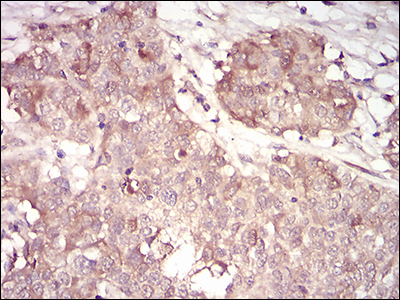
Immunohistochemical analysis of paraffin-embedded bladder cancer tissues using CCNA2 mouse mAb with DAB staining.
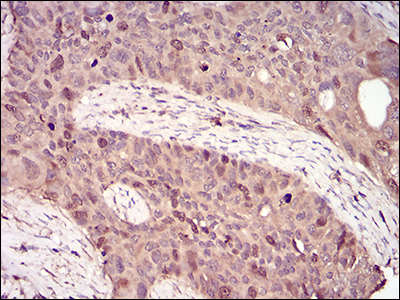
Immunohistochemical analysis of paraffin-embedded cervical cancer tissues using CCNA2 mouse mAb with DAB staining.
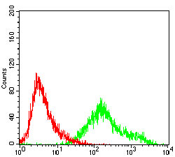
Flow cytometric analysis of A431 cells using CCNA2 mouse mAb (green) and negative control (red).
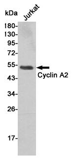
Western blot detection of Cyclin A2 in Jurkat cell lysates using Cyclin A2 mouse mAb (1:1000 diluted).Predicted band size:50KDa.Observed band size:50KDa.
-
Background
Swiss-Prot Acc.P20248.The protein encoded by this gene belongs to the highly conserved cyclin family, whose members are characterized by a dramatic periodicity in protein abundance through the cell cycle. Cyclins function as regulators of CDK kinases. Different cyclins exhibit distinct expression and degradation patterns which contribute to the temporal coordination of each mitotic event. In contrast to cyclin A1, which is present only in germ cells, this cyclin is expressed in all tissues tested. This cyclin binds and activates CDC2 or CDK2 kinases, and thus promotes both cell cycle G1/S and G2/M transitions.
Related Products / Services
Please note: All products are "FOR RESEARCH USE ONLY AND ARE NOT INTENDED FOR DIAGNOSTIC OR THERAPEUTIC USE"