
-
Product Name
Anti-CHI3L1 antibody
- Documents
-
Description
Mouse monoclonal antibody to CHI3L1
-
Tested applications
WB, IHC-P, ICC
-
Species reactivity
Human, Mouse
-
Alternative names
GP39 antibody; ASRT7 antibody; GP-39 antibody; YK-40 antibody; YKL40 antibody; CGP-39 antibody; YKL-40 antibody; YYL-40 antibody; HC-gp39 antibody; HCGP-3P antibody; hCGP-39 antibody
-
Isotype
Mouse IgG1
-
Preparation
This antigen of this antibody was synthetic peptide within human ykl-40 / chi3l1 aa 140-160.
-
Clonality
Monoclonal
-
Formulation
Liquid, 1*PBS (pH7.4), 0.2% BSA, 50% Glycerol. Preservative: 0.05% Sodium Azide.
-
Storage instructions
Store at +4℃ after thawing. Aliquot store at -20℃. Avoid repeated freeze / thaw cycles.
-
Applications
WB:1:500-1:1,000
IHC-P:1:50-1:200
FC:1:50-1:100
-
Validations
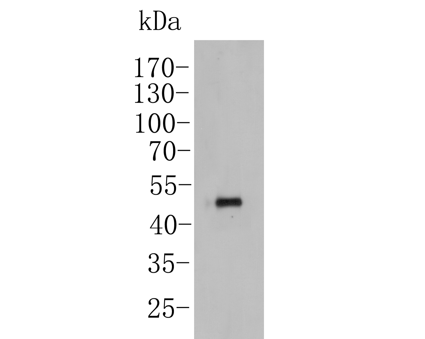
Fig1:; Western blot analysis of YKL-40 / CHI3L1 on THP-1 cell lysates. Proteins were transferred to a PVDF membrane and blocked with 5% BSA in PBS for 1 hour at room temperature. The primary antibody ( 1/500) was used in 5% BSA at room temperature for 2 hours. Goat Anti-Rabbit IgG - HRP Secondary Antibody (HA1001) at 1:5,000 dilution was used for 1 hour at room temperature.
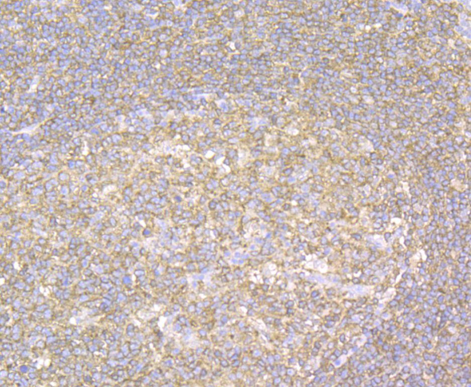
Fig2:; Immunohistochemical analysis of paraffin-embedded human tonsil tissue using anti-YKL-40 / CHI3L1 antibody. The section was pre-treated using heat mediated antigen retrieval with Tris-EDTA buffer (pH 8.0-8.4) for 20 minutes.The tissues were blocked in 5% BSA for 30 minutes at room temperature, washed with ddH; 2; O and PBS, and then probed with the antibody at 1/50 dilution, for 30 minutes at room temperature and detected using an HRP conjugated compact polymer system. DAB was used as the chrogen. Counter stained with hematoxylin and mounted with DPX.
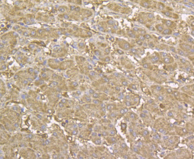
Fig3:; Immunohistochemical analysis of paraffin-embedded human liver tissue using anti-YKL-40 / CHI3L1 antibody. The section was pre-treated using heat mediated antigen retrieval with sodium citrate buffer (pH 6.0) for 20 minutes. The tissues were blocked in 5% BSA for 30 minutes at room temperature, washed with ddH; 2; O and PBS, and then probed with the antibody at 1/200 dilution, for 30 minutes at room temperature and detected using an HRP conjugated compact polymer system. DAB was used as the chrogen. Counter stained with hematoxylin and mounted with DPX.
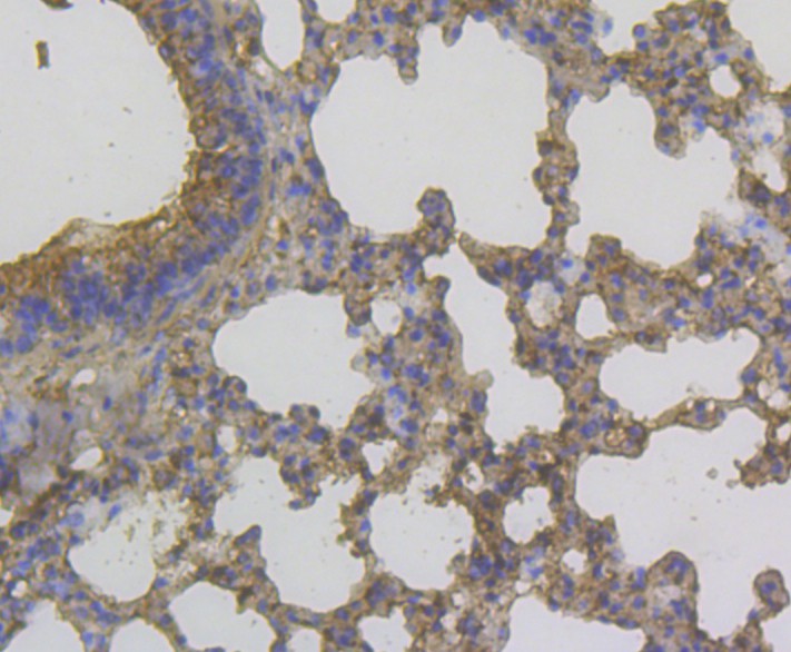
Fig4:; Immunohistochemical analysis of paraffin-embedded mouse lung tissue using anti-YKL-40 / CHI3L1 antibody. The section was pre-treated using heat mediated antigen retrieval with Tris-EDTA buffer (pH 8.0-8.4) for 20 minutes.The tissues were blocked in 5% BSA for 30 minutes at room temperature, washed with ddH; 2; O and PBS, and then probed with the antibody at 1/200 dilution, for 30 minutes at room temperature and detected using an HRP conjugated compact polymer system. DAB was used as the chrogen. Counter stained with hematoxylin and mounted with DPX.
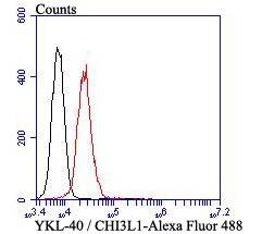
Fig5:; Flow cytometric analysis of YKL-40 / CHI3L1 was done on HepG2 cells. The cells were fixed, permeabilized and stained with YKL-40 / CHI3L1 antibody at 1/100 dilution (red) compared with an unlabelled control (cells without incubation with primary antibody; black). After incubation of the primary antibody on room temperature for an hour, the cells was stained with a Alexa Fluor™ 488-conjugated goat anti-mouse IgG Secondary antibody at 1/500 dilution for 30 minutes.
- Background
-
References
- Renkema G.H et al. Chitotriosidase, a chitinase, and the 39-kDa human cartilage glycoprotein, a chitin-binding lectin, are homologues of family 18 glycosyl hydrolases secreted by human macrophages. Eur J Biochem 251:504-509 (1998).
- Mizoguchi E. Chitinase 3-like-1 exacerbates intestinal inflammation by enhancing bacterial adhesion and invasion in colonic epithelial cells. Gastroenterology 130:398-411 (2006).
Related Products / Services
Please note: All products are "FOR RESEARCH USE ONLY AND ARE NOT INTENDED FOR DIAGNOSTIC OR THERAPEUTIC USE"