
-
Product Name
Anti-CCL2 antibody
- Documents
-
Description
Rabbit polyclonal antibody to CCL2
-
Tested applications
WB, IHC-P, ICC
-
Species reactivity
Human
-
Alternative names
HC11 antibody; MCAF antibody; MCP1 antibody; MCP-1 antibody; SCYA2 antibody; GDCF-2 antibody; SMC-CF antibody; HSMCR30 antibody
-
Isotype
Rabbit IgG
-
Preparation
This antigen of this antibody was synthetic peptide within human ccl2/mcp1 aa 50-99.
-
Clonality
Polyclonal
-
Formulation
Liquid, 1*TBS (pH7.4), 0.2% BSA, 50% Glycerol. Preservative: 0.05% Sodium Azide.
-
Storage instructions
Store at +4℃ after thawing. Aliquot store at -20℃. Avoid repeated freeze / thaw cycles.
-
Applications
WB: 1:500
IHC-P: 1:100-1:500
ICC: 1:50-1:200
-
Validations
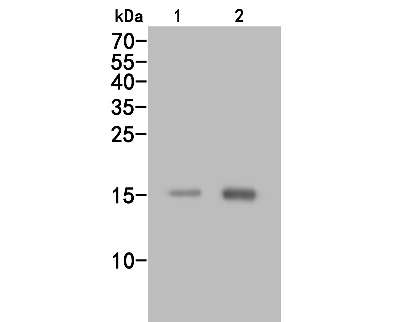
Fig1:; Western blot analysis of CCL2/MCP1 on different lysates. Proteins were transferred to a PVDF membrane and blocked with 5% BSA in PBS for 1 hour at room temperature. The primary antibody ( 1/500) was used in 5% BSA at room temperature for 2 hours. Goat Anti-Rabbit IgG - HRP Secondary Antibody (HA1001) at 1:5,000 dilution was used for 1 hour at room temperature.; Positive control:; Lane 1: A549 cell lysate; Lane 2: Hela cell lysate
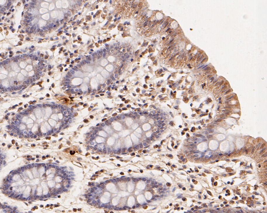
Fig2:; Immunohistochemical analysis of paraffin-embedded human colon tissue using anti-CCL2/MCP1 antibody. The section was pre-treated using heat mediated antigen retrieval with Tris-EDTA buffer (pH 8.0-8.4) for 20 minutes.The tissues were blocked in 5% BSA for 30 minutes at room temperature, washed with ddH; 2; O and PBS, and then probed with the primary antibody ( 1/800) for 30 minutes at room temperature. The detection was performed using an HRP conjugated compact polymer system. DAB was used as the chromogen. Tissues were counterstained with hematoxylin and mounted with DPX.
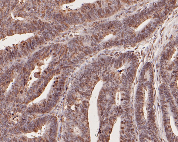
Fig3:; Immunohistochemical analysis of paraffin-embedded human colon carcinoma tissue using anti-CCL2/MCP1 antibody. The section was pre-treated using heat mediated antigen retrieval with Tris-EDTA buffer (pH 8.0-8.4) for 20 minutes.The tissues were blocked in 5% BSA for 30 minutes at room temperature, washed with ddH; 2; O and PBS, and then probed with the primary antibody ( 1/200) for 30 minutes at room temperature. The detection was performed using an HRP conjugated compact polymer system. DAB was used as the chromogen. Tissues were counterstained with hematoxylin and mounted with DPX.
- Background
-
References
- Li Y.S. et. al. The expression of monocyte chemotactic protein (MCP-1) in human vascular endothelium in vitro and in vivo. Mol. Cell. Biochem. 126:61-68(1993.
Related Products / Services
Please note: All products are "FOR RESEARCH USE ONLY AND ARE NOT INTENDED FOR DIAGNOSTIC OR THERAPEUTIC USE"