
-
Product Name
Anti-ATPB Rabbit antibody
- Documents
-
Description
ATPB Rabbit polyclonal antibody
-
Tested applications
WB, IHC-P, ICC/IF, IP
-
Species reactivity
Human, Mouse, Rat
-
Alternative names
ATP5B; ATPMB; ATPSB; HEL-S-271 antibody
-
Isotype
Rabbit IgG
-
Preparation
Antigen: A synthetic peptide of human ATPB
-
Clonality
Polyclonal
-
Formulation
Supplied in 50nM Tris-Glycine(pH 7.4), 0.15M Nacl, 40%Glycerol, 0.01% sodium azide and 0.05% BSA.
-
Storage instructions
Store at -20°C. Stable for 12 months from date of receipt.
-
Applications
WB: 1/2000-1/10000
IHC: 1/20-1/100
ICC/IF: 1/50
IP: 1/20-1/50
-
Validations
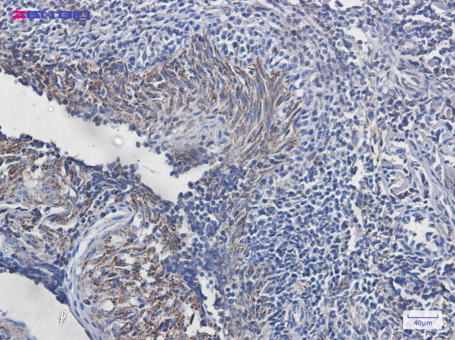
Immunohistochemistry of ATPB in paraffin-embedded Human tonsil using ATPB Rabbit pAb at dilution 1/20
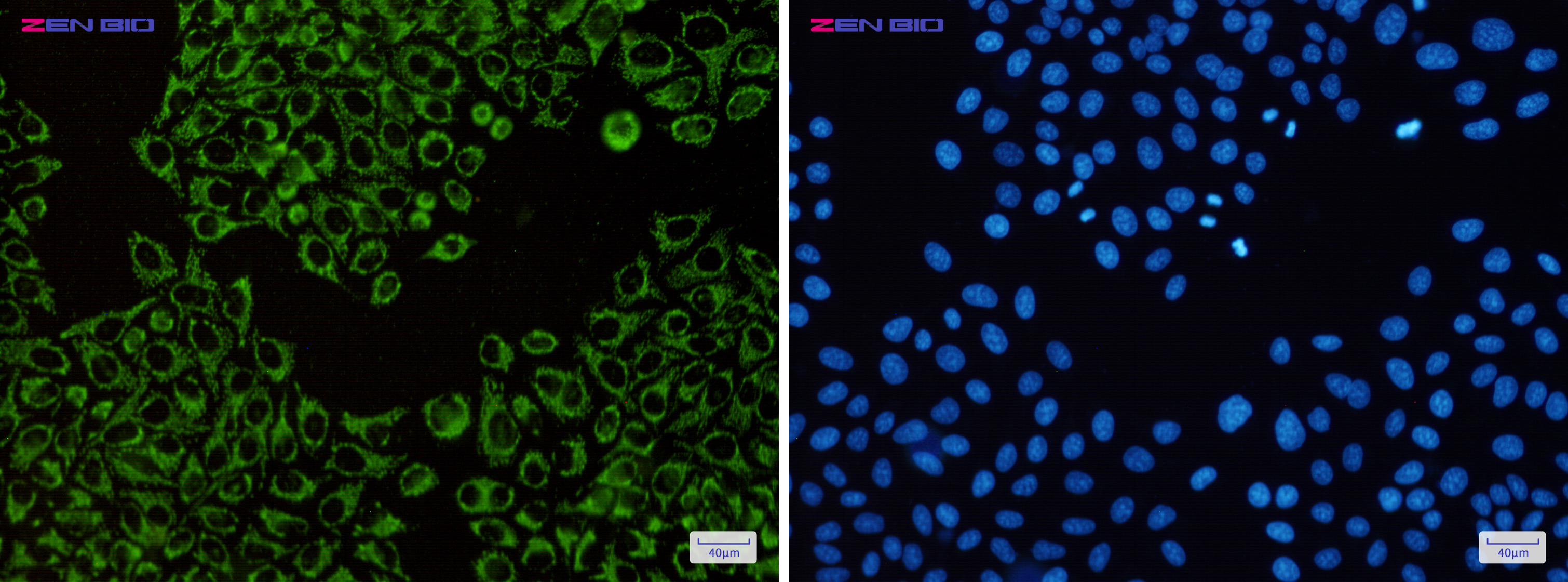
Immunocytochemistry of ATPB(green) in Hela cells using ATPB Rabbit pAb at dilution 1/50, and DAPI(blue)
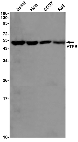
Western blot detection of ATPB in Jurkat,Hela,COS7,Raji cell lysates using ATPB Rabbit pAb(1:1000 diluted).Predicted band size:57KDa.Observed band size:52KDa.
-
Background
Swiss-Prot Acc.P06576.Mitochondrial membrane ATP synthase (F1F0 ATP synthase or Complex V) produces ATP from ADP in the presence of a proton gradient across the membrane which is generated by electron transport complexes of the respiratory chain. F-type ATPases consist of two structural domains, F1 - containing the extramembraneous catalytic core, and F0 - containing the membrane proton channel, linked together by a central stalk and a peripheral stalk. During catalysis, ATP synthesis in the catalytic domain of F1 is coupled via a rotary mechanism of the central stalk subunits to proton translocation. Subunits alpha and beta form the catalytic core in F1. Rotation of the central stalk against the surrounding alpha3beta3 subunits leads to hydrolysis of ATP in three separate catalytic sites on the beta subunits.
Related Products / Services
Please note: All products are "FOR RESEARCH USE ONLY AND ARE NOT INTENDED FOR DIAGNOSTIC OR THERAPEUTIC USE"