
-
Product Name
Anti-A2M antibody
- Documents
-
Description
Mouse monoclonal antibody to A2M
-
Tested applications
ELISA, ICC, IHC-P
-
Species reactivity
Human, Mouse, Rat
-
Alternative names
A2MD antibody; CPAMD5 antibody; FWP007 antibody; S863-7 antibody
-
Isotype
Mouse IgG1
-
Preparation
This antigen of this antibody was recombinant protein within human alpha-2-macroglobulin aa 950-1200.
-
Clonality
Monoclonal
-
Formulation
Liquid, 1*PBS (pH7.4), 0.2% BSA, 50% Glycerol. Preservative: 0.05% Sodium Azide.
-
Storage instructions
Store at +4℃ after thawing. Aliquot store at -20℃. Avoid repeated freeze / thaw cycles.
-
Applications
ELISA:1:1,000-1:5,000
WB:
IP:
ICC:1:50
IF:
IHC-P:1:50-1:200
FC:
-
Validations
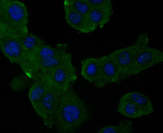
Fig1: ICC staining Alpha-2-macroglobulin in H22 cells (green). Formalin fixed cells were permeabilized with 0.1% Triton X-100 in TBS for 10 minutes at room temperature and blocked with 1% Blocker BSA for 15 minutes at room temperature. Cells were probed with Alpha-2-macroglobulin monoclonal antibody at a dilution of 1:50 for at least 1 hour at room temperature, washed with PBS. Alexa Fluorc™ 488 Goat anti-Mouse IgG was used as the secondary antibody at 1/100 dilution. The nuclear counter stain is DAPI (blue).
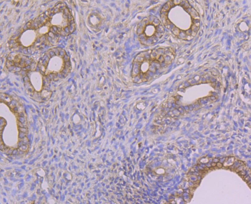
Fig2: Immunohistochemical analysis of paraffin-embedded rat uterus tissue using anti-Alpha-2-macroglobulin antibody. The section was pre-treated using heat mediated antigen retrieval with sodium citrate buffer (pH 6.0) for 20 minutes. The tissues were blocked in 5% BSA for 30 minutes at room temperature, washed with ddH2O and PBS, and then probed with the antibody at 1/50 dilution, for 30 minutes at room temperature and detected using an HRP conjugated compact polymer system. DAB was used as the chrogen. Counter stained with hematoxylin and mounted with DPX.
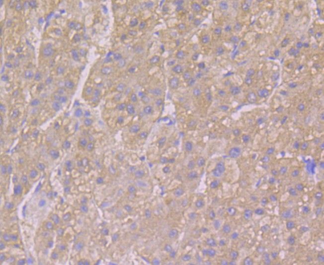
Fig3: Immunohistochemical analysis of paraffin-embedded human liver tissue using anti-Alpha-2-macroglobulin antibody. The section was pre-treated using heat mediated antigen retrieval with sodium citrate buffer (pH 6.0) for 20 minutes. The tissues were blocked in 5% BSA for 30 minutes at room temperature, washed with ddH2O and PBS, and then probed with the antibody at 1/50 dilution, for 30 minutes at room temperature and detected using an HRP conjugated compact polymer system. DAB was used as the chrogen. Counter stained with hematoxylin and mounted with DPX.
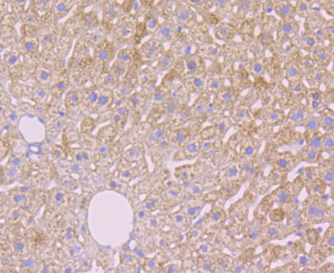
Fig4: Immunohistochemical analysis of paraffin-embedded mouse liver tissue using anti-Alpha-2-macroglobulin antibody. The section was pre-treated using heat mediated antigen retrieval with sodium citrate buffer (pH 6.0) for 20 minutes. The tissues were blocked in 5% BSA for 30 minutes at room temperature, washed with ddH2O and PBS, and then probed with the antibody at 1/50 dilution, for 30 minutes at room temperature and detected using an HRP conjugated compact polymer system. DAB was used as the chrogen. Counter stained with hematoxylin and mounted with DPX.
- Background
-
References
- Sottrup-Jensen L et al. Primary structure of human alpha 2-macroglobulin. V. The complete structure. J Biol Chem 259:8318-8327 (1984).
- Liu T et al. Human plasma N-glycoproteome analysis by immunoaffinity subtraction, hydrazide chemistry, and mass spectrometry. J Proteome Res 4:2070-2080 (2005).
Related Products / Services
Please note: All products are "FOR RESEARCH USE ONLY AND ARE NOT INTENDED FOR DIAGNOSTIC OR THERAPEUTIC USE"