
-
Product Name
MEGF10 Polyclonal Antibody
- Documents
-
Description
Polyclonal antibody to MEGF10
-
Tested applications
WB, IF
-
Species reactivity
Human, Mouse, Rat
-
Alternative names
MEGF10 antibody; EMARDD antibody; multiple EGF like domains 10 antibody
-
Isotype
Rabbit IgG
-
Preparation
Antigen: Recombinant fusion protein containing a sequence corresponding to amino acids 26-160 of human MEGF10 (NP_001295048.1).
-
Clonality
Polyclonal
-
Formulation
PBS with 0.02% sodium azide, 50% glycerol, pH7.3.
-
Storage instructions
Store at -20℃. Avoid freeze / thaw cycles.
-
Applications
WB 1:500 - 1:2000
IF 1:50 - 1:200 -
Validations
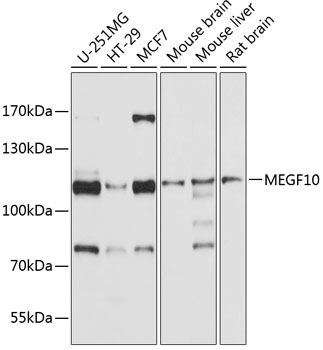
Western blot - MEGF10 Polyclonal Antibody
Western blot analysis of extracts of various cell lines, using MEGF10 antibody at 1:1000 dilution.Secondary antibody: HRP Goat Anti-Rabbit IgG (H+L) at 1:10000 dilution.Lysates/proteins: 25ug per lane.Blocking buffer: 3% nonfat dry milk in TBST.Detection: ECL Basic Kit .Exposure time: 10s.
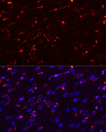
Immunofluorescence - MEGF10 Polyclonal Antibody
Immunofluorescence analysis of rat brain using MEGF10 antibody at dilution of 1:100. Blue: DAPI for nuclear staining.
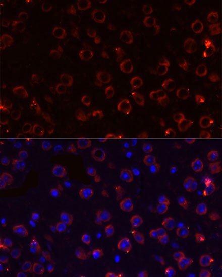
Immunofluorescence - MEGF10 Polyclonal Antibody
Immunofluorescence analysis of mouse brain using MEGF10 antibody at dilution of 1:100. Blue: DAPI for nuclear staining.
-
Background
Membrane receptor involved in phagocytosis by macrophages and astrocytes of apoptotic cells. Receptor for C1q, an eat-me signal, that binds phosphatidylserine expressed on the surface of apoptotic cells. Cooperates with ABCA1 within the process of engulfment. Promotes the formation of large intracellular vacuoles and may be responsible for the uptake of amyloid-beta peptides. Necessary for astrocyte-dependent apoptotic neuron clearance in the developing cerebellum. Plays role in muscle cell proliferation, adhesion and motility. Is also an essential factor in the regulation of myogenesis. Controls the balance between skeletal muscle satellite cells proliferation and differentiation through regulation of the notch signaling pathway. May also function in the mosaic spacing of specific neuron subtypes in the retina through homotypic retinal neuron repulsion. Mosaics provide a mechanism to distribute each cell type evenly across the retina, ensuring that all parts of the visual field have access to a full set of processing elements.
Related Products / Services
Please note: All products are "FOR RESEARCH USE ONLY AND ARE NOT INTENDED FOR DIAGNOSTIC OR THERAPEUTIC USE"