
-
Product Name
BIN1 antibody
- Documents
-
Description
BIN1 Rabbit Polyclonal antibody. Positive IP detected in mouse brain tissue. Positive WB detected in Jurkat cells, mouse brain tissue. Positive IHC detected in human osteosarcoma tissue. Observed molecular weight by Western-blot: 60-65 kDa
-
Tested applications
ELISA, WB, IHC, IP
-
Species reactivity
Human,Mouse,Rat; other species not tested.
-
Alternative names
AMPH2 antibody; Amphiphysin II antibody; Amphiphysin like protein antibody; AMPHL antibody; BIN1 antibody; bridging integrator 1 antibody; DKFZp547F068 antibody; SH3P9 antibody
-
Isotype
Rabbit IgG
-
Preparation
This antibody was obtained by immunization of BIN1 recombinant protein (Accession Number: NM_139350). Purification method: Antigen affinity purified.
-
Clonality
Polyclonal
-
Formulation
PBS with 0.02% sodium azide and 50% glycerol pH 7.3.
-
Storage instructions
Store at -20℃. DO NOT ALIQUOT
-
Applications
Recommended Dilution:
WB: 1:500-1:5000
IP: 1:200-1:2000
IHC: 1:20-1:200
-
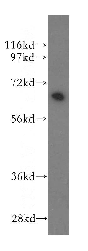
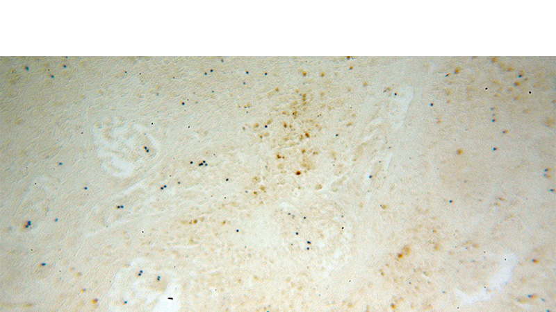
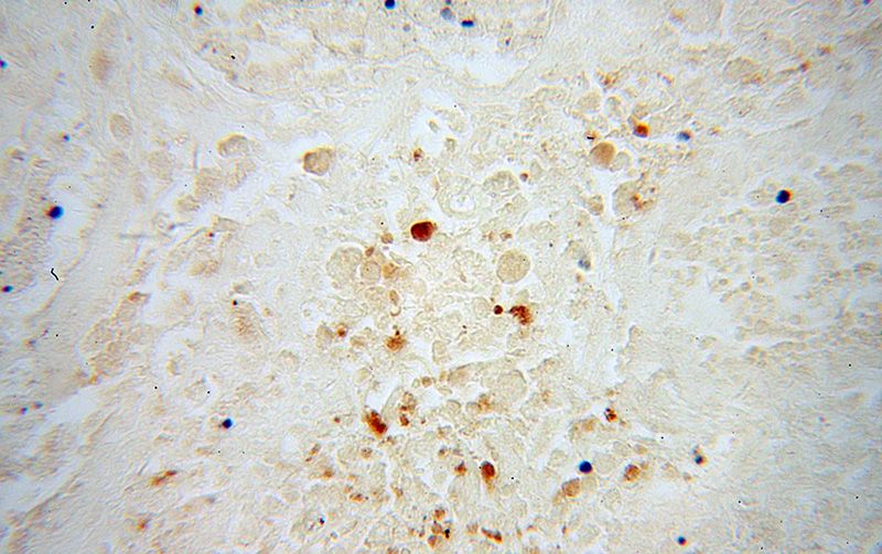
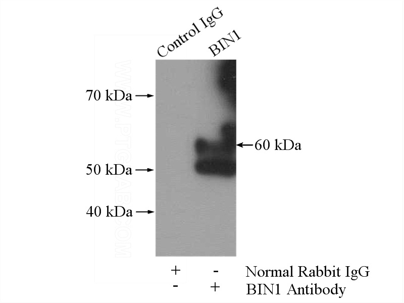
Validations
Jurkat cells were subjected to SDS PAGE followed by western blot with Catalog No:117146(BIN1 antibody) at dilution of 1:500
Immunohistochemical of paraffin-embedded human osteosarcoma using Catalog No:117146(BIN1 antibody) at dilution of 1:100 (under 10x lens)
Immunohistochemical of paraffin-embedded human osteosarcoma using Catalog No:117146(BIN1 antibody) at dilution of 1:100 (under 40x lens)
IP Result of anti-BIN1 (IP:Catalog No:117146, 4ug; Detection:Catalog No:117146 1:500) with mouse brain tissue lysate 3440ug.
-
Background
BIN1 (Bridging integrator 1), also known as amphiphysin II or Myc box-dependent-interacting protein 1, is a ubiquitous nucleocytoplasmic adaptor protein that was identified initially as an MYC-interacting proapoptotic tumor suppressor. Alternate splicing of the gene results in multiple transcript variants encoding different isoforms. BIN1 is a key regulator of different cellular functions, including endocytosis and membrane recycling, cytoskeleton regulation, DNA repair, cell cycle progression, and apoptosis (PMID: 24590001).
-
References
- Giridharan SS, Cai B, Vitale N, Naslavsky N, Caplan S. Cooperation of MICAL-L1, syndapin2, and phosphatidic acid in tubular recycling endosome biogenesis. Molecular biology of the cell. 24(11):1776-90, S1-15. 2013.
- Jia Y, Wang H, Wang Y. Low expression of Bin1, along with high expression of IDO in tumor tissue and draining lymph nodes, are predictors of poor prognosis for esophageal squamous cell cancer patients. International journal of cancer. 137(5):1095-106. 2015.
Related Products / Services
Please note: All products are "FOR RESEARCH USE ONLY AND ARE NOT INTENDED FOR DIAGNOSTIC OR THERAPEUTIC USE"