
-
Product Name
Anti-WSCD2 antibody
- Documents
-
Description
Mouse monoclonal antibody to WSCD2
-
Tested applications
WB, ICC, IHC-P
-
Species reactivity
Human
-
Isotype
Mouse IgM
-
Preparation
This antigen of this antibody was recombinant protein with human wscd2 50-353 / 565.
-
Clonality
Monoclonal
-
Formulation
Liquid, 1*PBS (pH7.4), 0.2% BSA, 50% Glycerol. Preservative: 0.05% Sodium Azide.
-
Storage instructions
Store at +4℃after thawing. Aliquot store at -20℃or -80℃Avoid repeated freeze / thaw cycles.
-
Applications
WB: 1:500-1:2,000
ICC: 1:50-1:200
IHC-P: 1:100-1:500
-
Validations
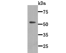
Fig1: Western blot analysis of WSCD2 on WSCD2 recombinant protein lysate using anti-WSCD2 antibody at 1/2000 dilution.
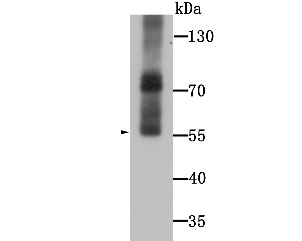
Fig2: Western blot analysis of WSCD2 on human stomach tissue lysate using anti-WSCD2 antibody at 1/1000 dilution.
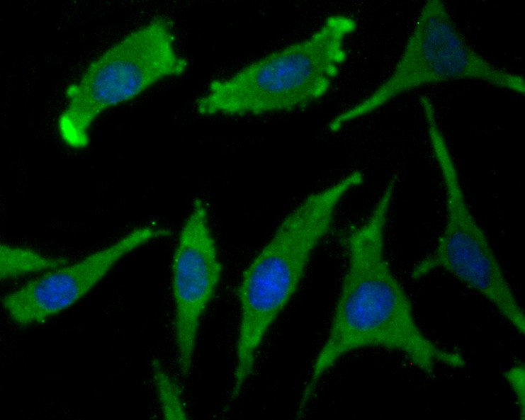
Fig3: ICC staining WSCD2 in SH-SY5Y cells (green). The nuclear counter stain is DAPI (blue). Cells were fixed in paraformaldehyde, permeabilised with 0.25% Triton X100/PBS.
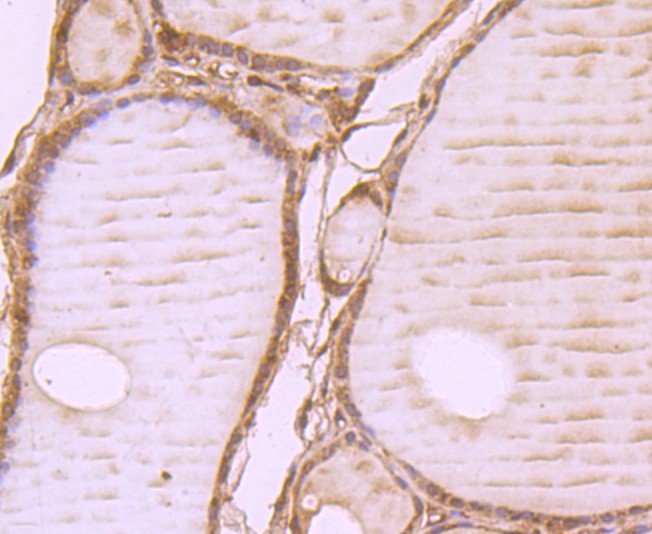
Fig4: Immunohistochemical analysis of paraffin-embedded human thyroid tissue using anti-WSCD2 antibody. Counter stained with
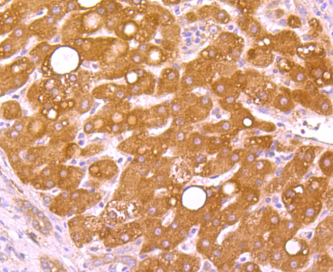
Fig5: Immunohistochemical analysis of paraffin-embedded human liver cancer tissue using anti-WSCD2 antibody. Counter stained with hematoxylin.
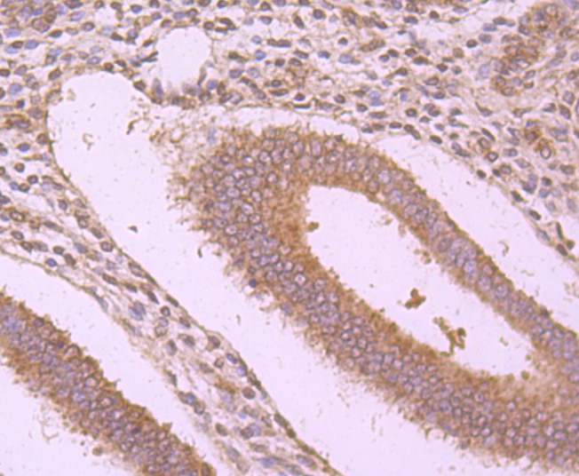
Fig6: Immunohistochemical analysis of paraffin-embedded human uterus tissue using anti-WSCD2 antibody. Counter stained with hematoxylin.
- Background
-
References
- Tong SM et al. Subcellular localization of five singular WSC domain-containing proteins and their roles in Beauveria bassiana responses to stress cues and metal ions. Environ Microbiol Rep 8(2):295-304 (2016).
- Gao C et al. A genome-wide linkage and association analysis of imputed insertions and deletions with cardiometabolic phenotypes in Mexican Americans: The Insulin Resistance Atherosclerosis Family Study. Genet Epidemiol 41(4):353-362 (2017).
Related Products / Services
Please note: All products are "FOR RESEARCH USE ONLY AND ARE NOT INTENDED FOR DIAGNOSTIC OR THERAPEUTIC USE"