
-
Product Name
Anti-TNFRSF9 antibody
- Documents
-
Description
Mouse monoclonal antibody to TNFRSF9
-
Tested applications
WB, IHC-P, ICC, FC
-
Species reactivity
Human, Mouse, Rat
-
Alternative names
ILA antibody; 4-1BB antibody; CD137 antibody; CDw137 antibody
-
Isotype
Mouse IgG1
-
Preparation
This antigen of this antibody was recombinant protein
-
Clonality
Monoclonal
-
Formulation
Liquid, 1*PBS (pH7.4), 0.2% BSA, 50% Glycerol. Preservative: 0.05% Sodium Azide.
-
Storage instructions
Store at +4℃ after thawing. Aliquot store at -20℃ or -80℃. Avoid repeated freeze / thaw cycles.
-
Applications
WB: 1:500
ICC: 1:100-1:500
IHC-P: 1:50-1:200
FC: 1:50-1:200
-
Validations
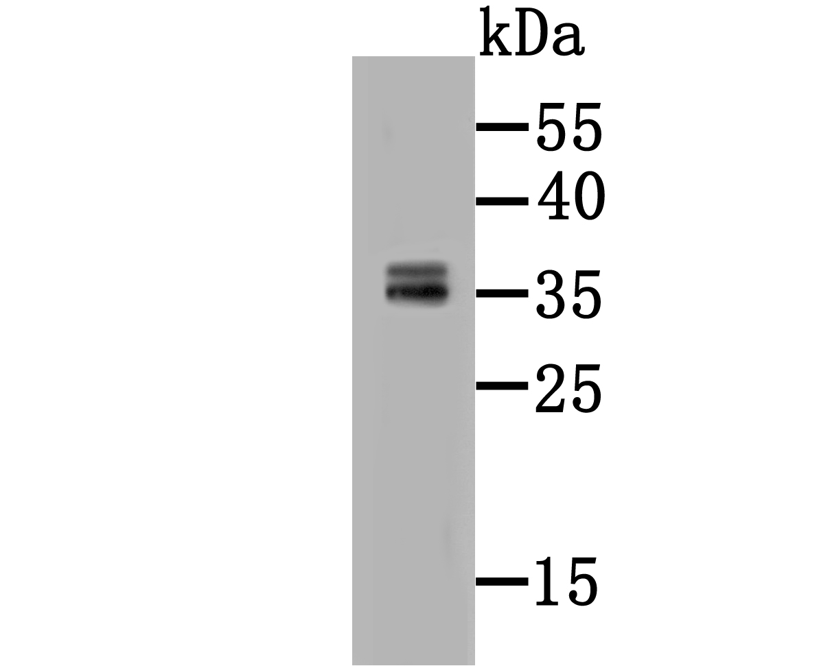
Fig1: Western blot analysis of CD137 on HepG2 cell lysate using anti-CD137 antibody at 1/500 dilution.
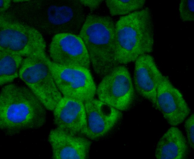
Fig2: ICC staining CD137 (green) in A431 cells. The nuclear counter stain is DAPI (blue). Cells were fixed in paraformaldehyde, permeabilised with 0.25% Triton X100/PBS.
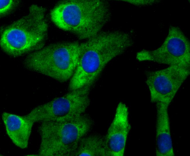
Fig3: ICC staining CD137 (green) in A549 cells. The nuclear counter stain is DAPI (blue). Cells were fixed in paraformaldehyde, permeabilised with 0.25% Triton X100/PBS.
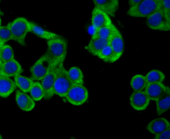
Fig4: ICC staining CD137 (green) in LOVO cells. The nuclear counter stain is DAPI (blue). Cells were fixed in paraformaldehyde, permeabilised with 0.25% Triton X100/PBS.
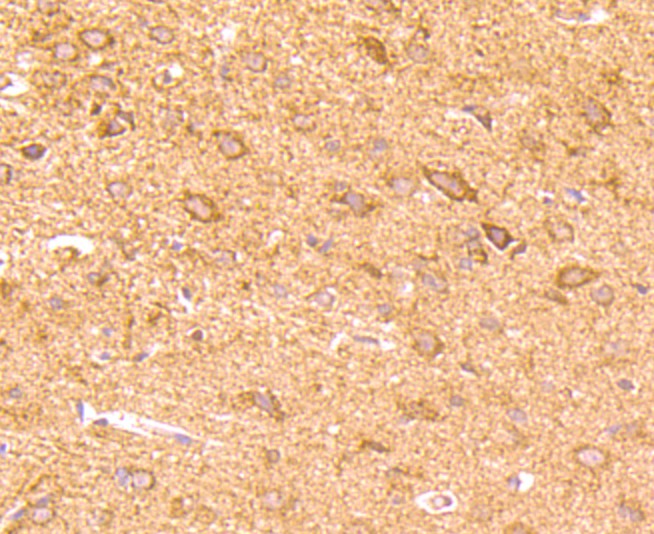
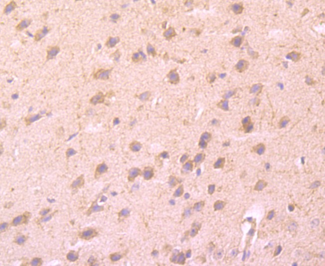
Fig5: Immunohistochemical analysis of paraffin-embedded rat brain tissue using anti-CD137 antibody. Counter stained with hematoxylin.
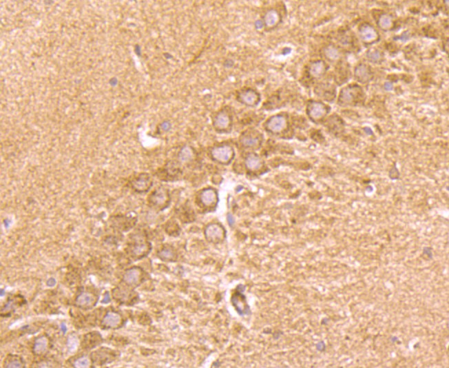
Fig6: Immunohistochemical analysis of paraffin-embedded rat hippocampus tissue using anti-CD137 antibody. Counter stained with hematoxylin.
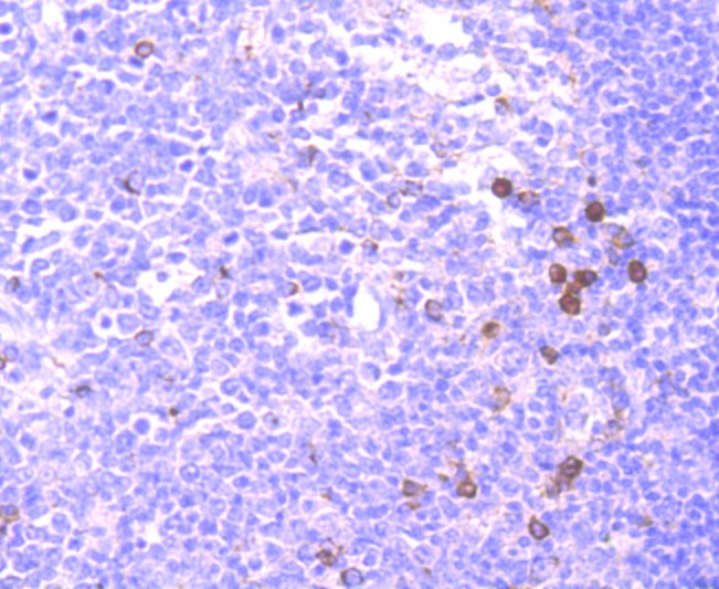
Fig7: Immunohistochemical analysis of paraffin-embedded human tonsil tissue using anti-CD137 antibody. Counter stained with hematoxylin.
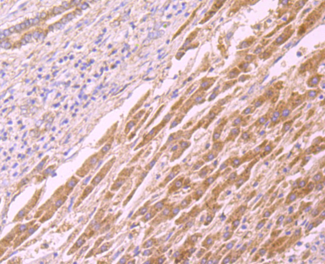
Fig8: Immunohistochemical analysis of paraffin-embedded human liver cancer tissue using anti-CD137 antibody. Counter stained with hematoxylin.
Fig9: Immunohistochemical analysis of paraffin-embedded mouse brain tissue using anti-CD137 antibody. Counter stained with hematoxylin.
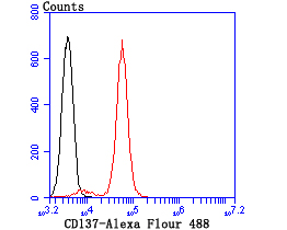
Fig10: Flow cytometric analysis of Jurkat cells with CD137 antibody at 1/100 dilution (red) compared with an unlabelled control (cells without incubation with primary antibody; black).
- Background
-
References
- Kang SW et al. Anti-CD137 suppresses tumor growth by blocking reverse signaling by CD137 ligand. Cancer Res. pii: canres (2017).
- Hebb JPO et al. Administration of low-dose combination anti-CTLA4, anti-CD137, and anti-OX40 into murine tumor or proximal to the tumor draining lymph node induces systemic tumor regression.Cancer Immunol Immunother.(2017).
Related Products / Services
Please note: All products are "FOR RESEARCH USE ONLY AND ARE NOT INTENDED FOR DIAGNOSTIC OR THERAPEUTIC USE"