
-
Product Name
Anti-TNFRSF19 antibody
- Documents
-
Description
Rabbit monoclonal antibody to TNFRSF19
-
Tested applications
WB, IHC-P, FC
-
Species reactivity
Human, Mouse, Rat
-
Alternative names
TAJ antibody; TROY antibody; TRADE antibody; TAJ-alpha antibody
-
Isotype
Rabbit IgG
-
Preparation
This antigen of this antibody was recombinant protein within human tnfrsf19 aa 50-200.
-
Clonality
Monoclonal
-
Formulation
Liquid, 1*TBS (pH7.4), 0.05% BSA, 40% Glycerol. Preservative: 0.05% Sodium Azide.
-
Storage instructions
Store at +4℃ after thawing. Aliquot store at -20℃. Avoid repeated freeze / thaw cycles.
-
Applications
WB:1:500-1:1,000
IHC-P:1:50-1:200
FC:1:50-1:100
-
Validations
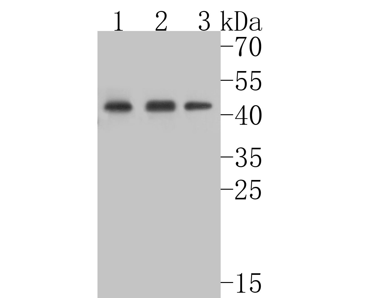
Fig1:; Western blot analysis of TNFRSF19 on different lysates. Proteins were transferred to a PVDF membrane and blocked with 5% BSA in PBS for 1 hour at room temperature. The primary antibody ( 1/500) was used in 5% BSA at room temperature for 2 hours. Goat Anti-Rabbit IgG - HRP Secondary Antibody (HA1001) at 1:5,000 dilution was used for 1 hour at room temperature.; Positive control:; Lane 1: mouse testis tissue lysate; Lane 2: rat large intestine tissue lysate; Lane 3: A431 cell lysate
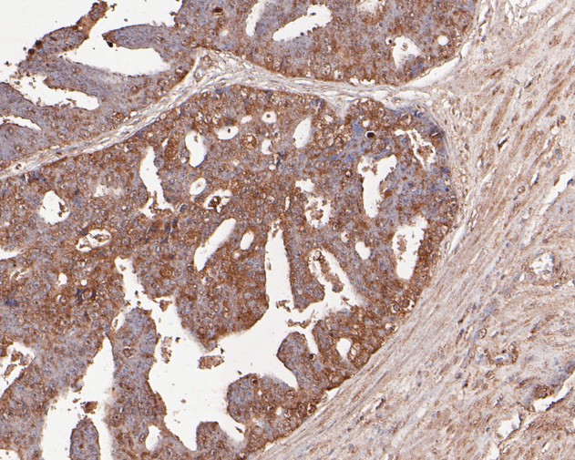
Fig2:; Immunohistochemical analysis of paraffin-embedded human prostate carcinoma tissue using anti-TNFRSF19 antibody. The section was pre-treated using heat mediated antigen retrieval with Tris-EDTA buffer (pH 8.0-8.4) for 20 minutes.The tissues were blocked in 5% BSA for 30 minutes at room temperature, washed with ddH; 2; O and PBS, and then probed with the primary antibody ( 1/200) for 30 minutes at room temperature. The detection was performed using an HRP conjugated compact polymer system. DAB was used as the chromogen. Tissues were counterstained with hematoxylin and mounted with DPX.
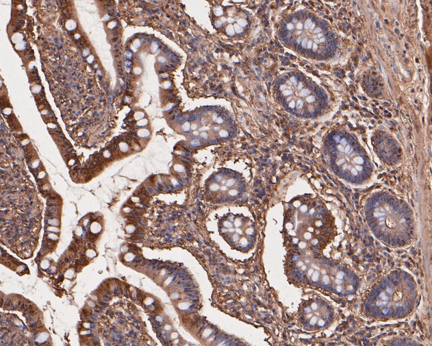
Fig3:; Immunohistochemical analysis of paraffin-embedded human small intestine tissue using anti-TNFRSF19 antibody. The section was pre-treated using heat mediated antigen retrieval with Tris-EDTA buffer (pH 8.0-8.4) for 20 minutes.The tissues were blocked in 5% BSA for 30 minutes at room temperature, washed with ddH; 2; O and PBS, and then probed with the primary antibody ( 1/50) for 30 minutes at room temperature. The detection was performed using an HRP conjugated compact polymer system. DAB was used as the chromogen. Tissues were counterstained with hematoxylin and mounted with DPX.
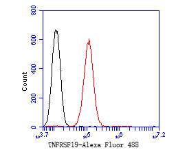
Fig4:; Flow cytometric analysis of TNFRSF19 was done on HepG2 cells. The cells were fixed, permeabilized and stained with the primary antibody ( 1/50) (red). After incubation of the primary antibody at room temperature for an hour, the cells were stained with a Alexa Fluor 488-conjugated Goat anti-Rabbit IgG Secondary antibody at 1/1000 dilution for 30 minutes.Unlabelled sample was used as a control (cells without incubation with primary antibody; black).
- Background
-
References
- Deng C. et. al. TNFRSF19 Inhibits TGFβ Signaling through Interaction with TGFβ Receptor Type I to Promote Tumorigenesis. Cancer Res. 2018 Jul.
- Shao L. et. al. The inherited variations of a p53-responsive enhancer in 13q12.12 confer lung cancer risk by attenuating TNFRSF19 expression. Genome Biol. 2019 May.
Related Products / Services
Please note: All products are "FOR RESEARCH USE ONLY AND ARE NOT INTENDED FOR DIAGNOSTIC OR THERAPEUTIC USE"