
-
Product Name
Anti-TNFRSF11B antibody
- Documents
-
Description
Mouse monoclonal antibody to TNFRSF11B
-
Tested applications
WB, ICC, IHC-P
-
Species reactivity
Human, Mouse, Rat
-
Alternative names
OPG antibody; TR1 antibody; OCIF antibody; PDB5 antibody
-
Isotype
Mouse IgG1
-
Preparation
This antigen of this antibody was recombinant protein within human osteoprotegerin aa 1-410.
-
Clonality
Monoclonal
-
Formulation
Liquid, 1*PBS (pH7.4), 0.2% BSA, 50% Glycerol. Preservative: 0.05% Sodium Azide.
-
Storage instructions
Store at +4℃ after thawing. Aliquot store at -20℃. Avoid repeated freeze / thaw cycles.
-
Applications
WB: 1:500-1:1,000
ICC: 1:50-1:200
IHC-P: 1:50-1:200
-
Validations
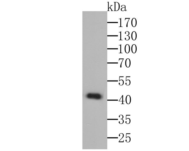
Fig1: Western blot analysis of Osteoprotegerin on K562 cell lysate using anti-Osteoprotegerin antibody at 1/1000 dilution.
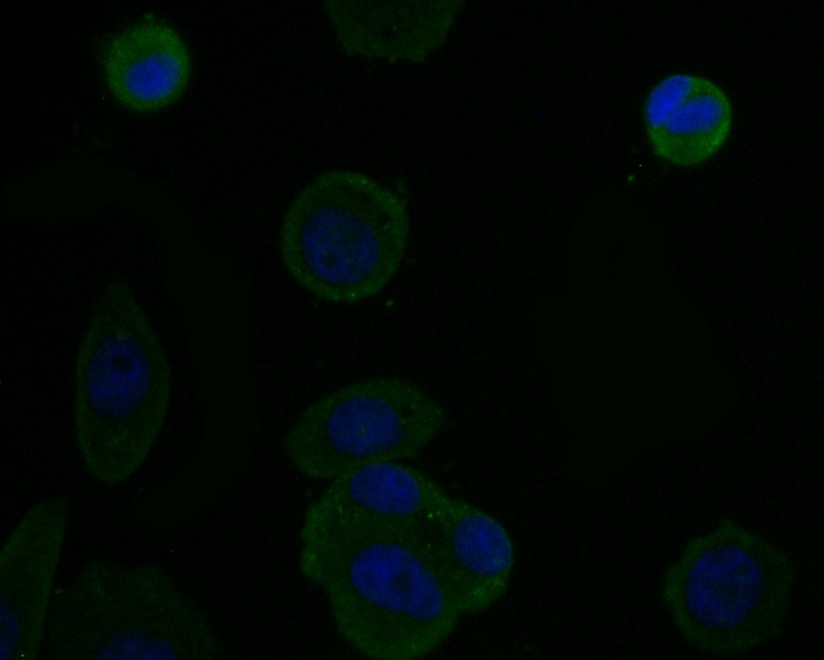
Fig2: ICC staining Osteoproteger in MCF-7 cells (green). The nuclear counter stain is DAPI (blue). Cells were fixed in paraformaldehyde, permeabilised with 0.25% Triton X100/PBS.
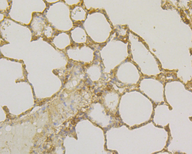
Fig3: Immunohistochemical analysis of paraffin-embedded rat lung tissue using anti-Osteoprotegerin antibody. Counter stained with hematoxylin.
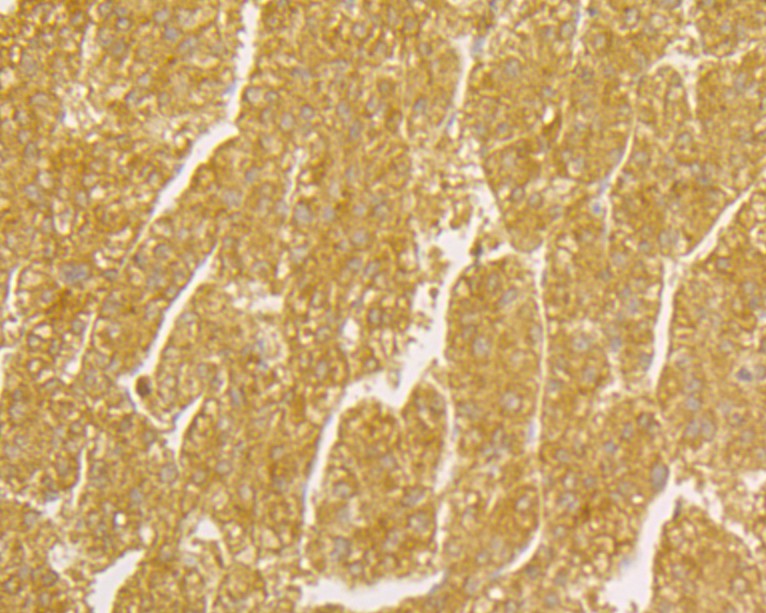
Fig4: Immunohistochemical analysis of paraffin-embedded human liver intestine tissue using anti-Osteoprotegerin antibody. Counter stained with hematoxylin.
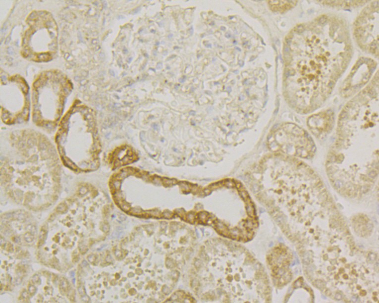
Fig5: Immunohistochemical analysis of paraffin-embedded human kidney tissue using anti-Osteoprotegerin antibody. Counter stained with hematoxylin.
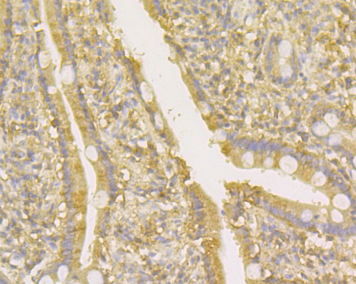
Fig6: Immunohistochemical analysis of paraffin-embedded human small intestine tissue using anti-Osteoprotegerin antibody. Counter stained with hematoxylin.
- Background
-
References
- Tsuda E et al. Isolation of a novel cytokine from human fibroblasts that specifically inhibits osteoclastogenesis. Biochem Biophys Res Commun 234:137-142 (1997).
- Luan X et al. Crystal structure of human RANKL complexed with its decoy receptor osteoprotegerin. J Immunol 189:245-252 (2012).
Related Products / Services
Please note: All products are "FOR RESEARCH USE ONLY AND ARE NOT INTENDED FOR DIAGNOSTIC OR THERAPEUTIC USE"