

-
Product Name
Anti-TMEM200A antibody
- Documents
-
Description
Mouse monoclonal antibody to TMEM200A
-
Tested applications
WB, ICC, IHC-P
-
Species reactivity
Human, Mouse
-
Alternative names
TTMA antibody; TTMC antibody; KIAA1913 antibody
-
Isotype
Mouse IgG1
-
Preparation
This antigen of this antibody was recombinant protein within human transmembrane protein 200a 50-400 aa.
-
Clonality
Monoclonal
-
Formulation
Liquid, 1*PBS (pH7.4), 0.2% BSA, 40% Glycerol. Preservative: 0.05% Sodium Azide.
-
Storage instructions
Store at +4℃ after thawing. Aliquot store at -20℃. Avoid repeated freeze / thaw cycles.
-
Applications
WB: 1:500
ICC: 1:100
IHC-P: 1:50
-
Validations
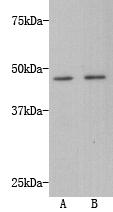
Fig1: Western blot analysis of Transmembrane protein 200A on A549 (1) and HepG2 (2) using anti-Transmembrane protein 200A antibody at 1/500 dilution.
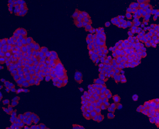
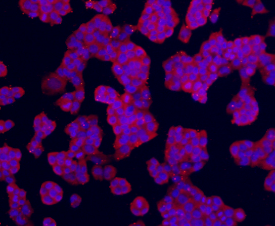
Fig2: ICC staining Transmembrane protein 200A (red) in F9 cells. The nuclear counter stain is DAPI (blue). Cells were fixed in paraformaldehyde, permeabilised with 0.25% Triton X100/PBS.
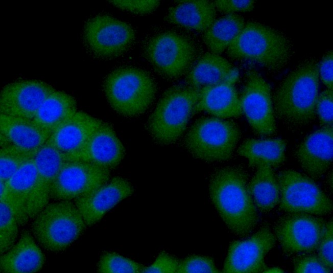
Fig3: ICC staining Transmembrane protein 200A (green) in HepG2 cells. The nuclear counter stain is DAPI (blue). Cells were fixed in paraformaldehyde, permeabilised with 0.25% Triton X100/PBS.
Fig4: ICC staining Transmembrane protein 200A (red) in NCCIT cells. The nuclear counter stain is DAPI (blue). Cells were fixed in paraformaldehyde, permeabilised with 0.25% Triton X100/PBS.
Fig5: Immunohistochemical analysis of paraffin-embedded human pancreas tissue using anti-Transmembrane protein 200A antibody. Counter stained with hematoxylin.
- Background
-
References
- Hui A B Y et al. Identification of a novel homozygous deletion region at 6q23.1 in medulloblastomas using high-resolution array comparative genomic hybridization analysis. Clin Cancer Res 11:4707-4716 (2005).
- Carninci P et al. The transcriptional landscape of the mammalian genome. Science 309:1559-1563 (2005).
Related Products / Services
Please note: All products are "FOR RESEARCH USE ONLY AND ARE NOT INTENDED FOR DIAGNOSTIC OR THERAPEUTIC USE"