
-
Product Name
Anti-Ripk3 antibody
- Documents
-
Description
Rabbit polyclonal antibody to Ripk3
-
Tested applications
WB
-
Species reactivity
Mouse, Rat
-
Alternative names
R antibody; Rip3 antibody; AW107945 antibody; 2610528K09Rik antibody
-
Isotype
Rabbit IgG
-
Preparation
This antigen of this antibody was synthetic peptide within mouse rip3 aa 440-486 / 486.
-
Clonality
Polyclonal
-
Formulation
Liquid, 1*PBS (pH7.4), 0.2% BSA, 50% Glycerol. Preservative: 0.05% Sodium Azide.
-
Storage instructions
Store at +4℃ after thawing. Aliquot store at -20℃. Avoid repeated freeze / thaw cycles.
-
Applications
WB: 1:500-1:1,000
-
Validations
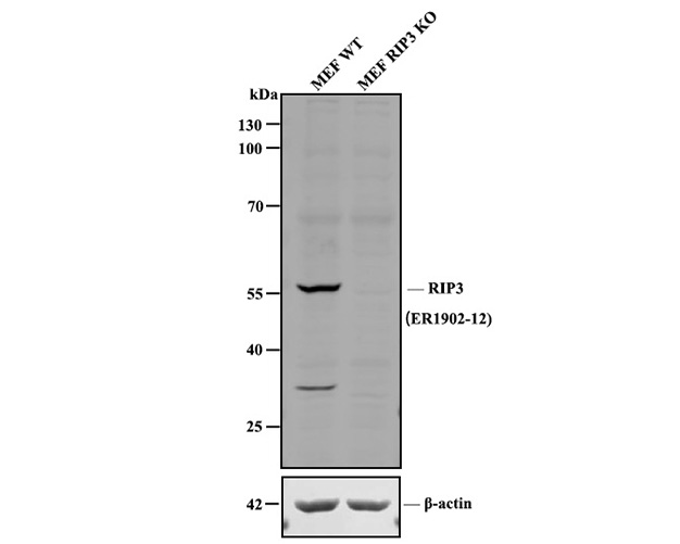
Fig1:; All lanes: Western blot analysis of RIP3 with anti-RIP3 antibody at 1:1,000 dilution.; Lane 1: Wild-type MEF whole cell lysate (20 µg).; Lane 2: RIP3 knockout MEF whole cell lysate (20 µg).; 175161# was shown to specifically react with RIP3 in wild-type MEF cells. No band was observed when RIP3 knockout sample was tested. Wild-type and RIP3 knockout samples were subjected to SDS-PAGE. Proteins were transferred to a PVDF membrane and blocked with 5% NFDM in TBST for 1 hour at room temperature. The primary antibody ( 1/1,000) and Loading control antibody (Rabbit anti-β-actin, R1207-1, 1/1,000) was used in 5% BSA at room temperature for 2 hours. Goat Anti-Rabbit IgG-HRP Secondary Antibody (HA1001) at 1:200,000 dilution was used for 1 hour at room temperature.
- Background
-
References
- Rebsamen M. et al, DAI/ZBP1 recruits RIP1 and RIP3 through RIP homotypic interaction motifs to activate NF-kappaB. EMBO Rep. 10:916-922(2009).
- Kaiser W.J. et al, Toll-like receptor 3-mediated necrosis via TRIF, RIP3, and MLKL. J. Biol. Chem. 288:31268-31279(2013).
Related Products / Services
Please note: All products are "FOR RESEARCH USE ONLY AND ARE NOT INTENDED FOR DIAGNOSTIC OR THERAPEUTIC USE"