

-
Product Name
Anti-PLEC antibody
- Documents
-
Description
Rabbit monoclonal antibody to PLEC
-
Tested applications
WB, IHC-P, IP
-
Species reactivity
Human, Mouse, Rat
-
Alternative names
HD1 antibody; PCN antibody; EBS1 antibody; EBSO antibody; PLTN antibody; EBSMD antibody; EBSND antibody; EBSOG antibody; EBSPA antibody; PLEC1 antibody; LGMD2Q antibody; PLEC1b antibody; LGMDR17 antibody
-
Isotype
Rabbit IgG
-
Preparation
This antigen of this antibody was recombinant protein within c-terminal human plectin.
-
Clonality
Monoclonal
-
Formulation
Liquid, 1*TBS (pH7.4), 0.05% BSA, 40% Glycerol. Preservative: 0.05% Sodium Azide.
-
Storage instructions
Store at +4℃ after thawing. Aliquot store at -20℃ or -80℃. Avoid repeated freeze / thaw cycles.
-
Applications
WB: 1:1,000
IHC-P: 1:100-1:500
-
Validations
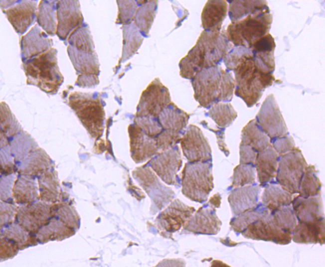
Fig1:; Immunohistochemical analysis of paraffin-embedded rat skeletal muscle tissue using anti-Plectin antibody. The section was pre-treated using heat mediated antigen retrieval with Tris-EDTA buffer (pH 8.0-8.4) for 20 minutes.The tissues were blocked in 5% BSA for 30 minutes at room temperature, washed with ddH; 2; O and PBS, and then probed with the primary antibody ( 1/50) for 30 minutes at room temperature. The detection was performed using an HRP conjugated compact polymer system. DAB was used as the chromogen. Tissues were counterstained with hematoxylin and mounted with DPX.
Fig2:; Immunohistochemical analysis of paraffin-embedded mouse skeletal muscle tissue using anti-Plectin antibody. The section was pre-treated using heat mediated antigen retrieval with Tris-EDTA buffer (pH 8.0-8.4) for 20 minutes.The tissues were blocked in 5% BSA for 30 minutes at room temperature, washed with ddH; 2; O and PBS, and then probed with the primary antibody ( 1/50) for 30 minutes at room temperature. The detection was performed using an HRP conjugated compact polymer system. DAB was used as the chromogen. Tissues were counterstained with hematoxylin and mounted with DPX.
- Background
-
References
- Kulkarni RM et al. Ron receptor signaling is protective against DSS-induced colitis in mice. Am J Physiol Gastrointest Liver Physiol 306:G1065-74 (2014).
- Eisenberg JL et al. Plectin-containing, centrally localized focal adhesions exert traction forces in primary lung epithelial cells. J Cell Sci 126:3746-55 (2013).
Related Products / Services
Please note: All products are "FOR RESEARCH USE ONLY AND ARE NOT INTENDED FOR DIAGNOSTIC OR THERAPEUTIC USE"