
-
Product Name
Anti-PKC eta Rabbit antibody
- Documents
-
Description
PKC eta Rabbit monoclonal antibody
-
Tested applications
WB, IP
-
Species reactivity
Human, Mouse, Rat
-
Isotype
Rabbit IgG
-
Preparation
Antigen: A synthetic peptide of human PKC eta
-
Clonality
Monoclonal
-
Formulation
Supplied in 50nM Tris-Glycine(pH 7.4), 0.15M Nacl, 40%Glycerol, 0.01% sodium azide and 0.05% BSA.
-
Storage instructions
Store at 4°C short term. Aliquot and store at -20°C long term. Avoid freeze / thaw cycle.
-
Applications
1:1000
1:20
-
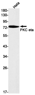
Validations
Western blot detection of PKC eta in Hela cell lysates using PKC eta Rabbit mAb(1:1000 diluted).Predicted band size:78kDa.Observed band size:78kDa.
-
Background
Swiss-Prot Acc.P24723.Calcium-independent, phospholipid- and diacylglycerol (DAG)-dependent serine/threonine-protein kinase that is involved in the regulation of cell differentiation in keratinocytes and pre-B cell receptor, mediates regulation of epithelial tight junction integrity and foam cell formation, and is required for glioblastoma proliferation and apoptosis prevention in MCF-7 cells. In keratinocytes, binds and activates the tyrosine kinase FYN, which in turn blocks epidermal growth factor receptor (EGFR) signaling and leads to keratinocyte growth arrest and differentiation. Associates with the cyclin CCNE1-CDK2-CDKN1B complex and inhibits CDK2 kinase activity, leading to RB1 dephosphorylation and thereby G1 arrest in keratinocytes. In association with RALA activates actin depolymerization, which is necessary for keratinocyte differentiation. In the pre-B cell receptor signaling, functions downstream of BLNK by up-regulating IRF4, which in turn activates L chain gene rearrangement. Regulates epithelial tight junctions (TJs) by phosphorylating occludin (OCLN) on threonine residues, which is necessary for the assembly and maintenance of TJs. In association with PLD2 and via TLR4 signaling, is involved in lipopolysaccharide (LPS)-induced RGS2 down-regulation and foam cell formation. Upon PMA stimulation, mediates glioblastoma cell proliferation by activating the mTOR pathway, the PI3K/AKT pathway and the ERK1-dependent phosphorylation of ELK1. Involved in the protection of glioblastoma cells from irradiation-induced apoptosis by preventing caspase-9 activation. In camptothecin-treated MCF-7 cells, regulates NF-kappa-B upstream signaling by activating IKBKB, and confers protection against DNA damage-induced apoptosis. Promotes oncogenic functions of ATF2 in the nucleus while blocking its apoptotic function at mitochondria. Phosphorylates ATF2 which promotes its nuclear retention and transcriptional activity and negatively regulates its mitochondrial localization.
Related Products / Services
Please note: All products are "FOR RESEARCH USE ONLY AND ARE NOT INTENDED FOR DIAGNOSTIC OR THERAPEUTIC USE"