

-
Product Name
Anti-OSBPL1A antibody
- Documents
-
Description
Rabbit monoclonal antibody to OSBPL1A
-
Tested applications
WB, IHC-P
-
Species reactivity
Human, Mouse, Rat
-
Alternative names
ORP1 antibody; ORP-1 antibody; OSBPL1B antibody
-
Isotype
Rabbit IgG
-
Preparation
This antigen of this antibody was recombinant protein.
-
Clonality
Monoclonal
-
Formulation
Liquid, 1*TBS (pH7.4), 0.05% BSA, 40% Glycerol. Preservative: 0.05% Sodium Azide.
-
Storage instructions
Store at +4℃ after thawing. Aliquot store at -20℃. Avoid repeated freeze / thaw cycles.
-
Applications
WB: 1:500-1:2,000
IHC-P: 1:50-1:200
-
Validations
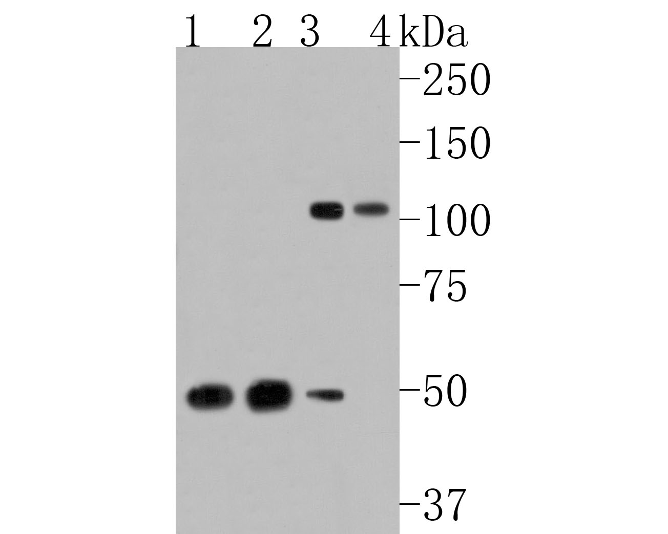
Fig1:; Western blot analysis of ORP1 on different lysates. Proteins were transferred to a PVDF membrane and blocked with 5% NFTM/TBST for 1 hour at room temperature. The primary antibody ( 1/500) was used in 5% NFTM/TBST at room temperature for 2 hours. Goat Anti-Rabbit IgG - HRP Secondary Antibody (HA1001) at 1:200,000 dilution was used for 1 hour at room temperature.; Positive control:; Lane 1: Mouse heart tissue lysate; Lane 2: Rat brain tissue lysate; Lane 3: A549 cell lysate; Lane 4: PC-3 cell lysate
Fig2:; Immunohistochemical analysis of paraffin-embedded mouse skeletal muscle tissue using anti-ORP1 antibody. The section was pre-treated using heat mediated antigen retrieval with Tris-EDTA buffer (pH 9.0) for 20 minutes.The tissues were blocked in 5% BSA for 30 minutes at room temperature, washed with ddH; 2; O and PBS, and then probed with the primary antibody ( 1/50) for 30 minutes at room temperature. The detection was performed using an HRP conjugated compact polymer system. DAB was used as the chromogen. Tissues were counterstained with hematoxylin and mounted with DPX.
- Background
-
References
- Tong J. et. al. Structural basis of human ORP1-Rab7 interaction for the late-endosome and lysosome targeting. PLoS One. 2019 Feb
- Dong J. et. al. Allosteric enhancement of ORP1-mediated cholesterol transport by PI(4,5)P(2)/PI(3,4)P(2). Nat Commun. 2019 Feb
Related Products / Services
Please note: All products are "FOR RESEARCH USE ONLY AND ARE NOT INTENDED FOR DIAGNOSTIC OR THERAPEUTIC USE"