
-
Product Name
Anti-NTRK1 antibody
- Documents
-
Description
Rabbit polyclonal antibody to NTRK1
-
Tested applications
IHC-P, FC, WB, ICC
-
Species reactivity
Human, Mouse, Rat
-
Alternative names
MTC antibody; TRK antibody; TRK1 antibody; TRKA antibody; Trk-A antibody; p140-TrkA antibody
-
Isotype
Rabbit IgG
-
Preparation
This antigen of this antibody was recombinant protein
-
Clonality
Polyclonal
-
Formulation
Liquid, 1*PBS (pH7.4), 0.2% BSA, 50% Glycerol. Preservative: 0.05% Sodium Azide.
-
Storage instructions
Store at +4℃ after thawing. Aliquot store at -20℃ or -80℃. Avoid repeated freeze / thaw cycles.
-
Applications
WB: 1:500
ICC: 1:100
IHC-P: 1:50-1:200
FC: 1:50-1:100
-
Validations
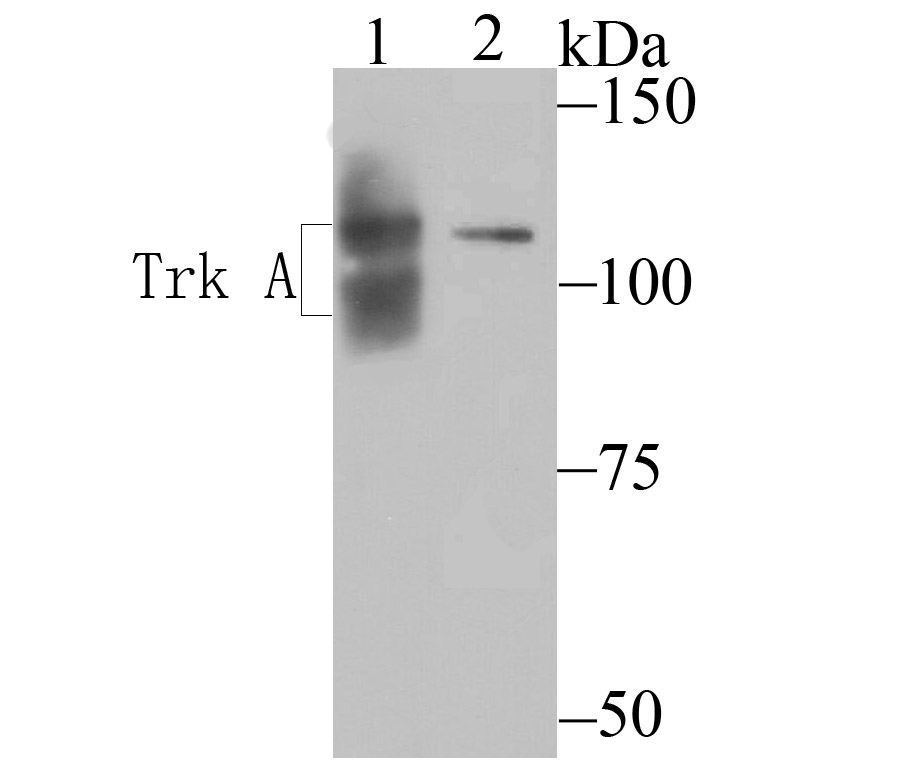
Fig1: Western blot analysis of TrkA on SHSY5Y (1) and SHG-44 (2) cell lysates using anti-TrkA antibody at 1/200 dilution.
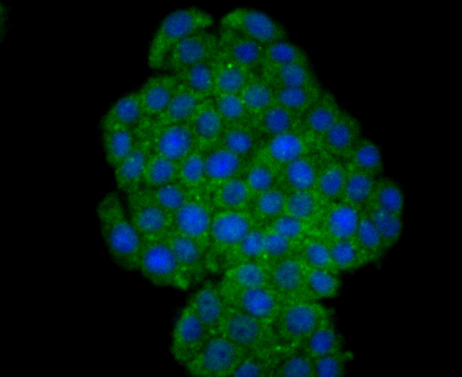
Fig2: ICC staining TrkA in PC-12 cells (green). The nuclear counter stain is DAPI (blue). Cells were fixed in paraformaldehyde, permeabilised with 0.25% Triton X100/PBS.
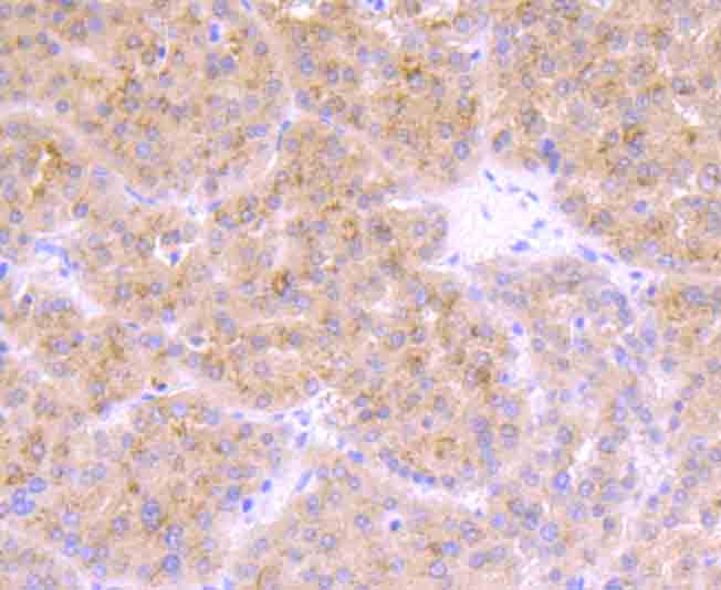
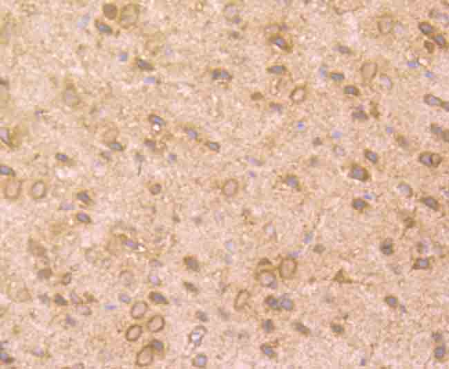
Fig3: Immunohistochemical analysis of paraffin-embedded human liver cancer tissue using anti-TrkA antibody. Counter stained with hematoxylin.
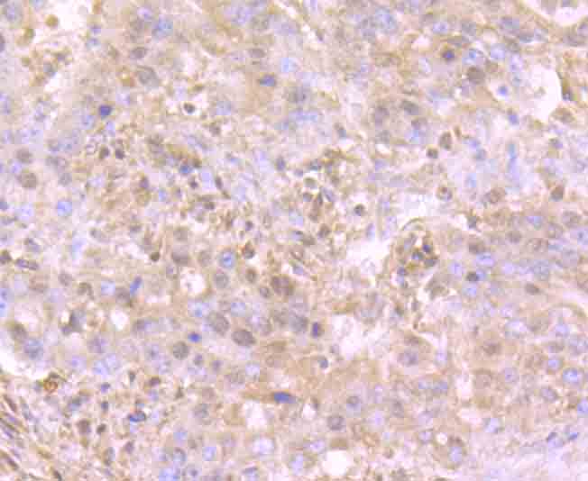
Fig4: Immunohistochemical analysis of paraffin-embedded human stomach cancer tissue using anti-TrkA antibody. Counter stained with hematoxylin.
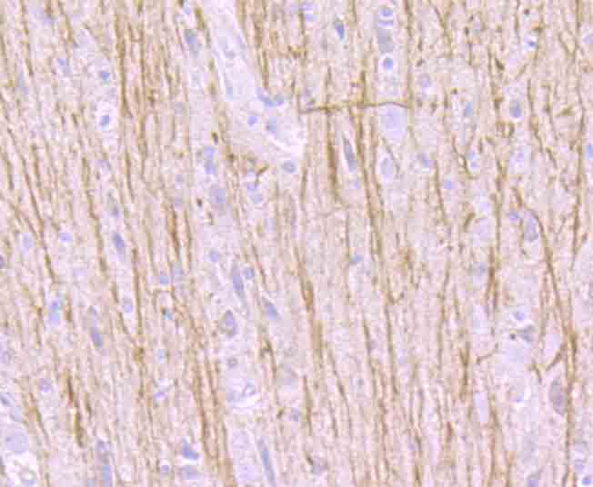
Fig5: Immunohistochemical analysis of paraffin-embedded rat brain tissue using anti-TrkA antibody. Counter stained with hematoxylin.
Fig6: Immunohistochemical analysis of paraffin-embedded mouse brain tissue using anti-TrkA antibody. Counter stained with hematoxylin.
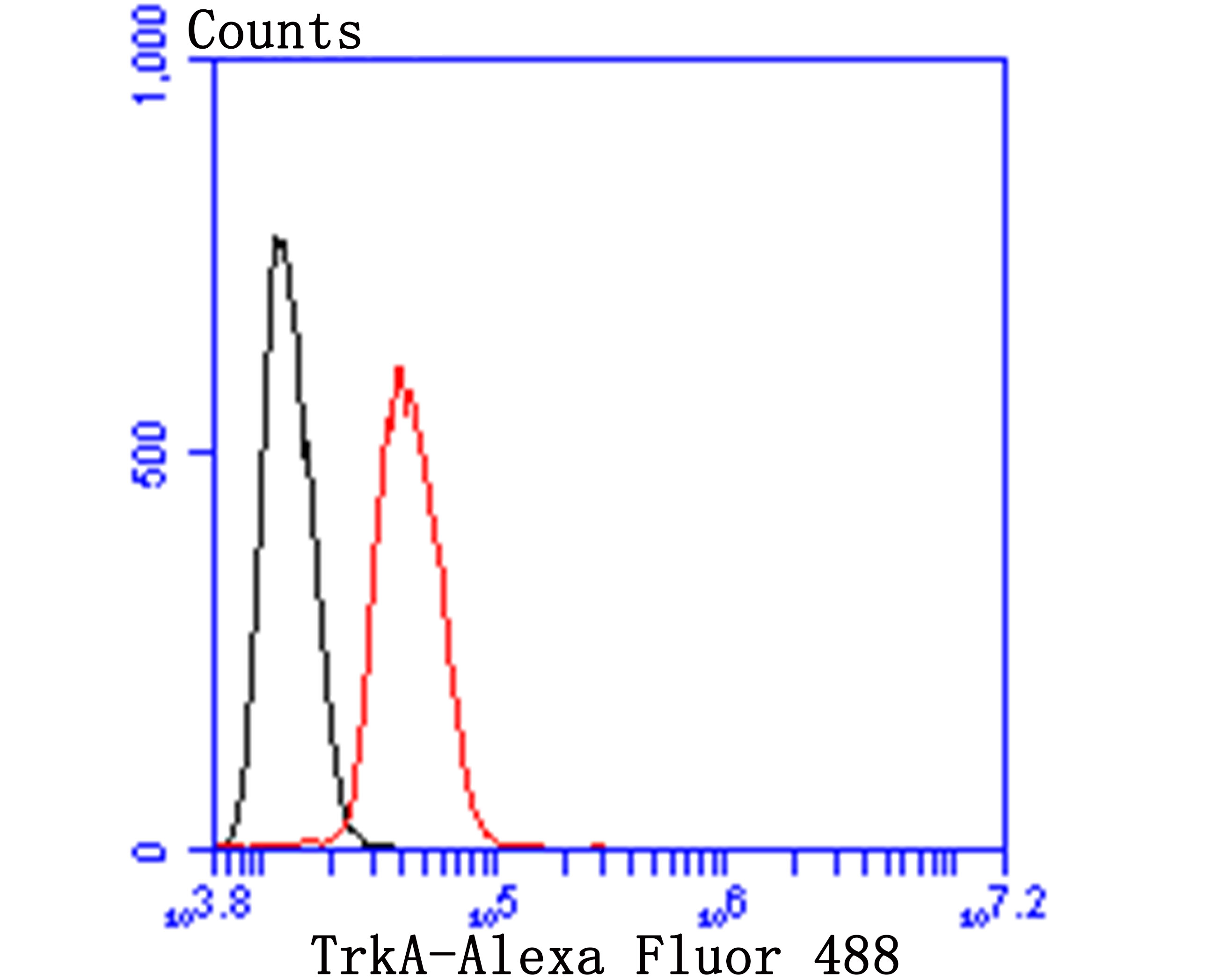
Fig7: Flow cytometric analysis of SHSY5Y cells with TrkA 1/2 antibody at 1/100 dilution (blue) compared with an unlabelled control (cells without incubation with primary antibody; red). Goat anti rabbit IgG (FITC) was used as the secondary antibody.
- Background
-
References
- Hempstead B L et al. High-affinity NGF binding requires coexpression of the trk proto-oncogene and the low-affinity NGF receptor. Nature 350:678-683 (1991).
- Jing S et al. Nerve growth factor mediates signal transduction through trk homodimer receptors. Neuron 9:1067-1079 (1992).
Related Products / Services
Please note: All products are "FOR RESEARCH USE ONLY AND ARE NOT INTENDED FOR DIAGNOSTIC OR THERAPEUTIC USE"