
-
Product Name
Anti-NPY5R antibody
- Documents
-
Description
Rabbit monoclonal antibody to NPY5R
-
Tested applications
WB, ICC, IHC-P
-
Species reactivity
Human, Mouse, Rat
-
Alternative names
NPYR5 antibody; NPY5-R antibody; NPYY5-R antibody
-
Isotype
Rabbit IgG
-
Preparation
This antigen of this antibody was recombinant protein.
-
Clonality
Monoclonal
-
Formulation
Liquid, 1*TBS (pH7.4), 0.05% BSA, 40% Glycerol. Preservative: 0.05% Sodium Azide.
-
Storage instructions
Store at +4℃ after thawing. Aliquot store at -20℃. Avoid repeated freeze / thaw cycles.
-
Applications
WB: 1:500-1:2,000
ICC: 1:50-1:200
IHC-P: 1:50-1:200
-
Validations
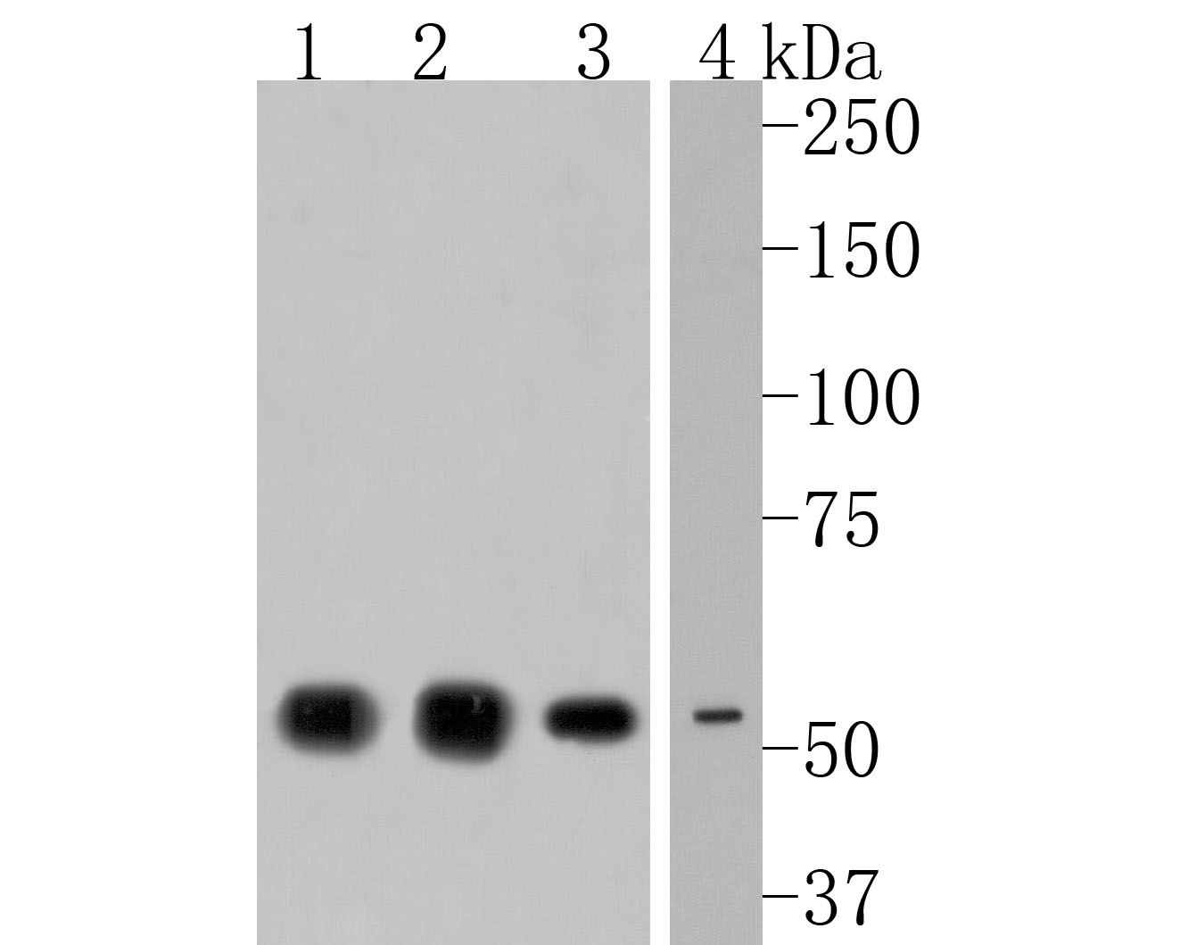
Fig1:; Western blot analysis of NPY5R on different lysates. Proteins were transferred to a PVDF membrane and blocked with 5% NFTM/TBST for 1 hour at room temperature. The primary antibody ( 1/500) was used in 5% NFTM/TBST at room temperature for 2 hours. Goat Anti-Rabbit IgG - HRP Secondary Antibody (HA1001) at 1:200,000 dilution was used for 1 hour at room temperature.; Positive control:; Lane 1: Human brain tissue lysate; Lane 2: Rat brain tissue lysate; Lane 3: A549 cell lysate; Lane 4: Mouse spleen tissue lysate
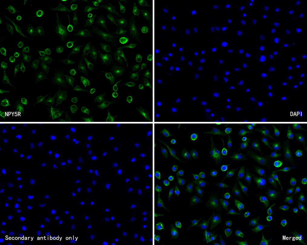
Fig2:; ICC staining of NPY5R in A549 cells (green). Formalin fixed cells were permeabilized with 0.1% Triton X-100 in TBS for 10 minutes at room temperature and blocked with 1% Blocker BSA for 15 minutes at room temperature. Cells were probed with the primary antibody ( 1/200) for 1 hour at room temperature, washed with PBS. Alexa Fluor®488 Goat anti-Rabbit IgG was used as the secondary antibody at 1/1,000 dilution. The nuclear counter stain is DAPI (blue).
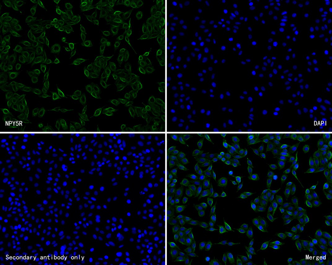
Fig3:; ICC staining of NPY5R in SiHa cells (green). Formalin fixed cells were permeabilized with 0.1% Triton X-100 in TBS for 10 minutes at room temperature and blocked with 1% Blocker BSA for 15 minutes at room temperature. Cells were probed with the primary antibody ( 1/50) for 1 hour at room temperature, washed with PBS. Alexa Fluor®488 Goat anti-Rabbit IgG was used as the secondary antibody at 1/1,000 dilution. The nuclear counter stain is DAPI (blue).
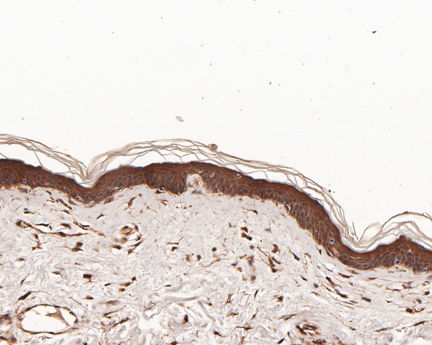
Fig4:; Immunohistochemical analysis of paraffin-embedded human skin tissue using anti-NPY5R antibody. The section was pre-treated using heat mediated antigen retrieval with Tris-EDTA buffer (pH 9.0) for 20 minutes.The tissues were blocked in 5% BSA for 30 minutes at room temperature, washed with ddH; 2; O and PBS, and then probed with the primary antibody ( 1/50) for 30 minutes at room temperature. The detection was performed using an HRP conjugated compact polymer system. DAB was used as the chromogen. Tissues were counterstained with hematoxylin and mounted with DPX.
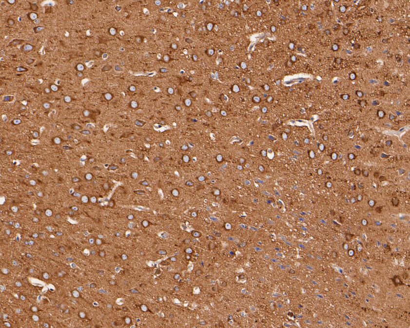
Fig5:; Immunohistochemical analysis of paraffin-embedded mouse brain tissue using anti-NPY5R antibody. The section was pre-treated using heat mediated antigen retrieval with Tris-EDTA buffer (pH 9.0) for 20 minutes.The tissues were blocked in 5% BSA for 30 minutes at room temperature, washed with ddH; 2; O and PBS, and then probed with the primary antibody ( 1/50) for 30 minutes at room temperature. The detection was performed using an HRP conjugated compact polymer system. DAB was used as the chromogen. Tissues were counterstained with hematoxylin and mounted with DPX.
- Background
-
References
- Kumar JS. et. al. Radiosynthesis and in Vivo Evaluation of Neuropeptide Y5 Receptor (NPY5R) PET Tracers. ACS Chem Neurosci. 2016 May
- Erondu N. et. al. NPY5R antagonism does not augment the weight loss efficacy of orlistat or sibutramine. Obesity (Silver Spring). 2007 Aug
Related Products / Services
Please note: All products are "FOR RESEARCH USE ONLY AND ARE NOT INTENDED FOR DIAGNOSTIC OR THERAPEUTIC USE"