
-
Product Name
Anti-MYT1L antibody
- Documents
-
Description
Rabbit polyclonal antibody to MYT1L
-
Tested applications
WB, IHC, FC
-
Species reactivity
Human, Mouse
-
Alternative names
NZF1 antibody; MRD39 antibody; myT1-L antibody; ZC2H2C2 antibody; ZC2HC4B antibody
-
Isotype
Rabbit IgG
-
Preparation
This antigen of this antibody was recombinant protein within human myt1l aa 200-400 / 1186.
-
Clonality
Polyclonal
-
Formulation
Liquid, 1*TBS (pH7.4), 0.2% BSA, 50% Glycerol. Preservative: 0.05% Sodium Azide.
-
Storage instructions
Store at +4℃ after thawing. Aliquot store at -20℃. Avoid repeated freeze / thaw cycles.
-
Applications
WB: 1:500-1:1000
IHC: 1:100-1:500
FC: 1:100-1:500
-
Validations
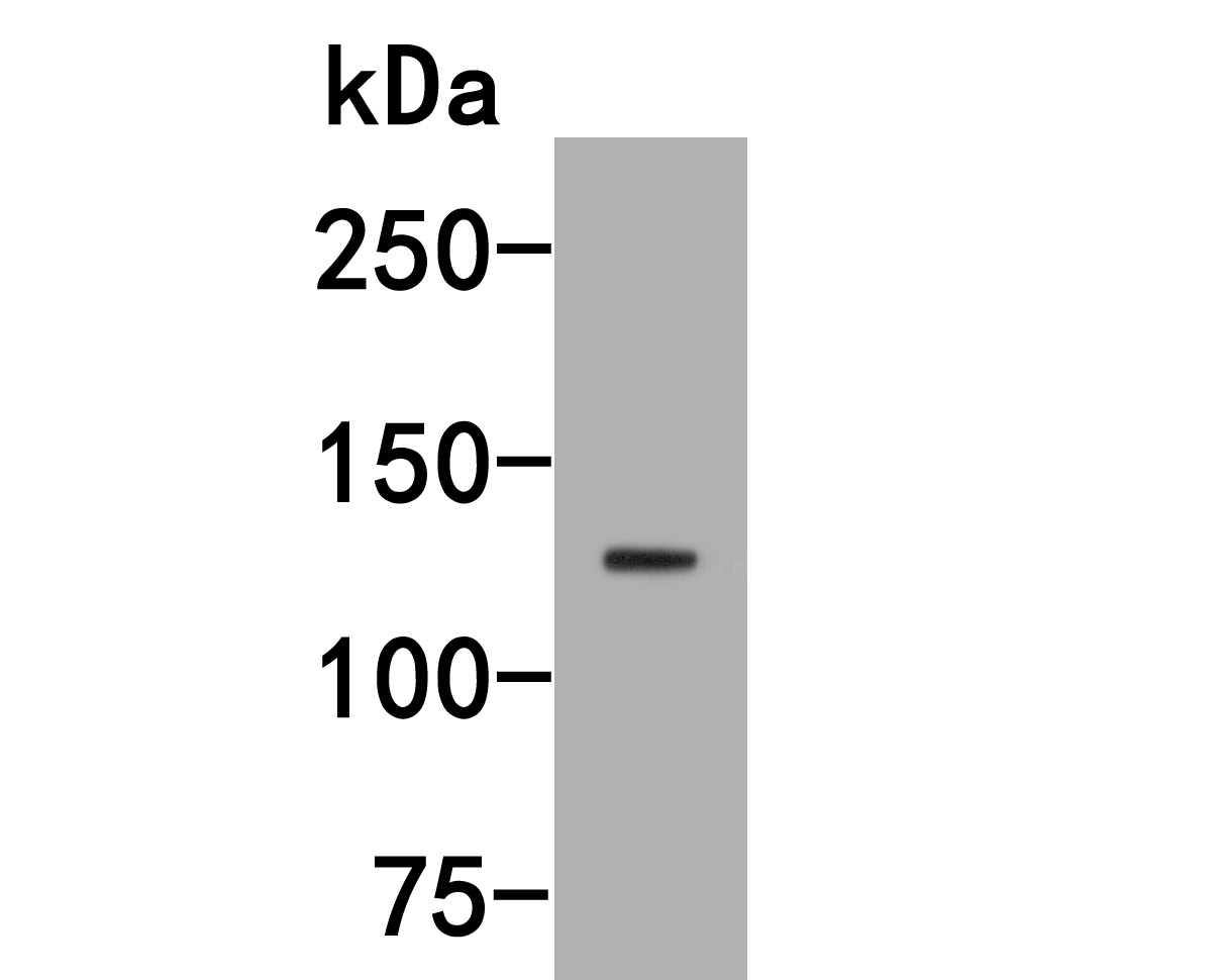
Fig1:; Western blot analysis of MyT1L on SHSY5Y cell lysates. Proteins were transferred to a PVDF membrane and blocked with 5% NFDM/TBST for 1 hour at room temperature. The primary antibody ( 1/1,000) was used in 5% NFDM/TBST at room temperature for 2 hours. Goat Anti-Rabbit IgG - HRP Secondary Antibody (HA1001) at 1:200,000 dilution was used for 1 hour at room temperature.
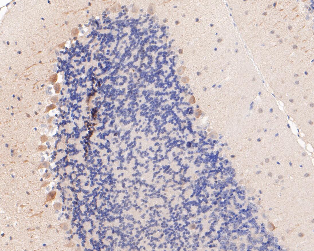
Fig2:; Immunohistochemical analysis of paraffin-embedded mouse cerebellum tissue using anti-MyT1L antibody. The section was pre-treated using heat mediated antigen retrieval with sodium citrate buffer (pH 6.0) for 20 minutes. The tissues were blocked in 5% BSA for 30 minutes at room temperature, washed with ddH; 2; O and PBS, and then probed with the primary antibody ( 1/400) for 30 minutes at room temperature. The detection was performed using an HRP conjugated compact polymer system. DAB was used as the chromogen. Tissues were counterstained with hematoxylin and mounted with DPX.
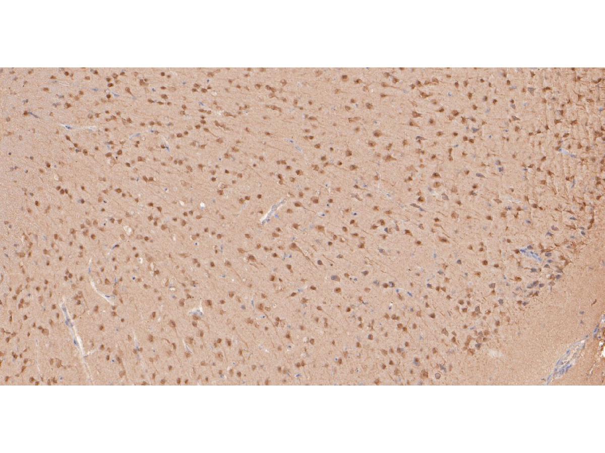
Fig3:; Immunohistochemical analysis of paraffin-embedded mouse brain tissue using anti-MyT1L antibody. The section was pre-treated using heat mediated antigen retrieval with sodium citrate buffer (pH 6.0) for 20 minutes. The tissues were blocked in 5% BSA for 30 minutes at room temperature, washed with ddH; 2; O and PBS, and then probed with the primary antibody ( 1/400) for 30 minutes at room temperature. The detection was performed using an HRP conjugated compact polymer system. DAB was used as the chromogen. Tissues were counterstained with hematoxylin and mounted with DPX.
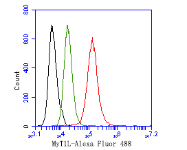
Fig4:; Flow cytometric analysis of MyT1L was done on SH-SY5Y cells. The cells were fixed, permeabilized and stained with the primary antibody ( 1ug/ml) (red) compared with Rabbit IgG, monoclonal - Isotype Control (green). After incubation of the primary antibody at +4℃ for 1 hour, the cells were stained with a Alexa Fluor®488 conjugate-Goat anti-Rabbit IgG Secondary antibody at 1/1,000 dilution for 30 minutes at +4℃ (dark incubation).Unlabelled sample was used as a control (cells without incubation with primary antibody; black).
- Background
-
References
- de Ligt J. et. al. Diagnostic exome sequencing in persons with severe intellectual disability. N. Engl. J. Med. 367:1921-1929(2012).
Related Products / Services
Please note: All products are "FOR RESEARCH USE ONLY AND ARE NOT INTENDED FOR DIAGNOSTIC OR THERAPEUTIC USE"