
-
Product Name
Anti-KCNK18 antibody
- Documents
-
Description
Rabbit polyclonal antibody to KCNK18
-
Tested applications
WB, ICC, IHC-P, FC
-
Species reactivity
Human, Mouse, Rat
-
Alternative names
TRIK antibody; MGR13 antibody; TRESK antibody; TRESK2 antibody; K2p18.1 antibody; TRESK-2 antibody
-
Isotype
Rabbit IgG
-
Preparation
This antigen of this antibody was synthetic peptide within mouse kcnk18 aa 44-93 / 394.
-
Clonality
Polyclonal
-
Formulation
Liquid, 1*PBS (pH7.4), 0.2% BSA, 50% Glycerol. Preservative: 0.05% Sodium Azide.
-
Storage instructions
Store at +4℃ after thawing. Aliquot store at -20℃. Avoid repeated freeze / thaw cycles.
-
Applications
WB:1:500-1:2,000
ICC:1:50-1:100
IHC-P:1:100-1:500
FC:1:100-1:500
-
Validations
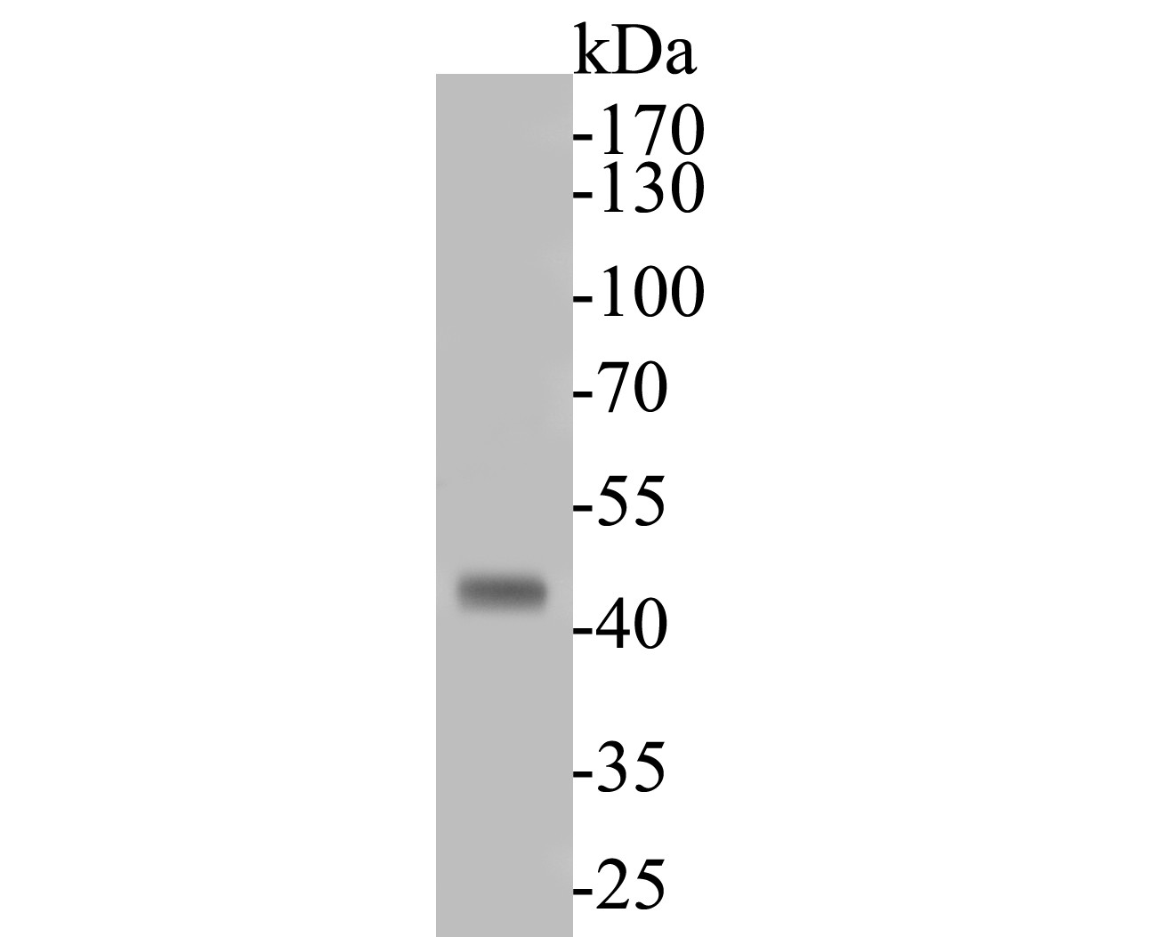
Fig1: Western blot analysis of KCNK18 on rat spinal cord tissue lysates. Proteins were transferred to a PVDF membrane and blocked with 5% BSA in PBS for 1 hour at room temperature. The primary antibody ( 1/200) was used in 5% BSA at room temperature for 2 hours. Goat Anti-Rabbit IgG - HRP Secondary Antibody (HA1001) at 1:5,000 dilution was used for 1 hour at room temperature.
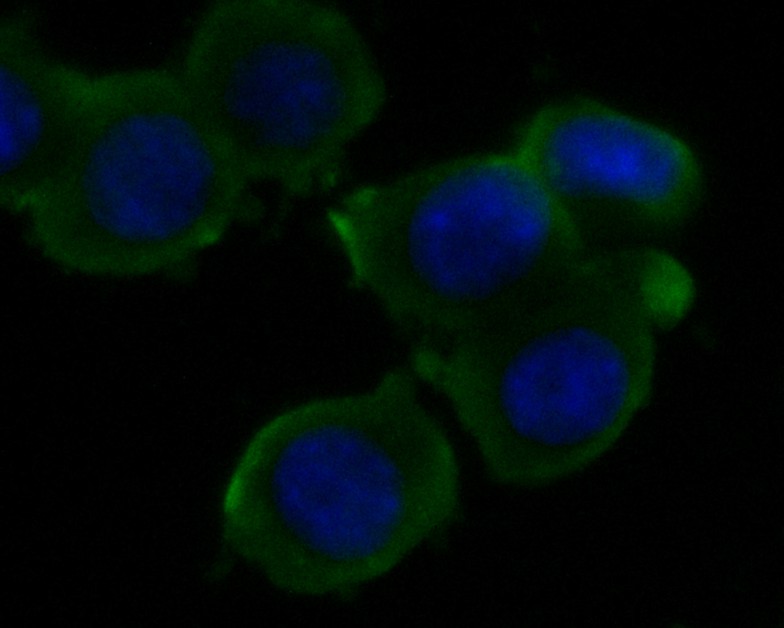
Fig2: ICC staining of KCNK18 in N2A cells (green). Formalin fixed cells were permeabilized with 0.1% Triton X-100 in TBS for 10 minutes at room temperature and blocked with 1% Blocker BSA for 15 minutes at room temperature. Cells were probed with the primary antibody ( 1/50) for 1 hour at room temperature, washed with PBS. Alexa Fluor®488 Goat anti-Rabbit IgG was used as the secondary antibody at 1/1,000 dilution. The nuclear counter stain is DAPI (blue).
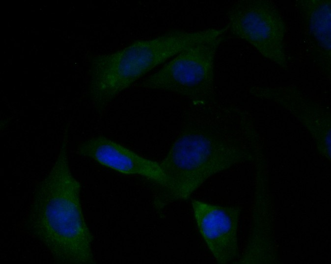
Fig3: ICC staining of KCNK18 in SHG-44 cells (green). Formalin fixed cells were permeabilized with 0.1% Triton X-100 in TBS for 10 minutes at room temperature and blocked with 1% Blocker BSA for 15 minutes at room temperature. Cells were probed with the primary antibody ( 1/100) for 1 hour at room temperature, washed with PBS. Alexa Fluor®488 Goat anti-Rabbit IgG was used as the secondary antibody at 1/1,000 dilution. The nuclear counter stain is DAPI (blue).
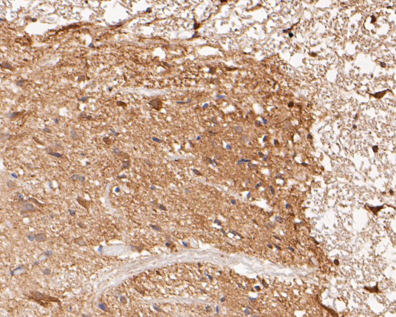
Fig4: Immunohistochemical analysis of paraffin-embedded rat spinal cord tissue using anti-KCNK18 antibody. The section was pre-treated using heat mediated antigen retrieval with Tris-EDTA buffer (pH 8.0-8.4) for 20 minutes.The tissues were blocked in 5% BSA for 30 minutes at room temperature, washed with ddH2O and PBS, and then probed with the primary antibody ( 1/50) for 30 minutes at room temperature. The detection was performed using an HRP conjugated compact polymer system. DAB was used as the chromogen. Tissues were counterstained with hematoxylin and mounted with DPX.
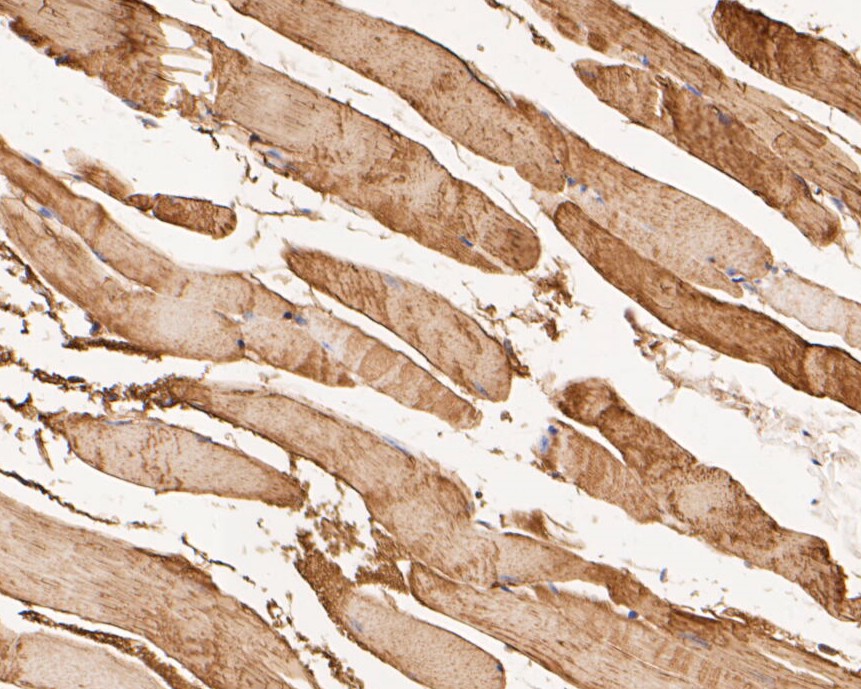
Fig5: Immunohistochemical analysis of paraffin-embedded mouse skeletal muscle tissue using anti-KCNK18 antibody. The section was pre-treated using heat mediated antigen retrieval with Tris-EDTA buffer (pH 8.0-8.4) for 20 minutes.The tissues were blocked in 5% BSA for 30 minutes at room temperature, washed with ddH2O and PBS, and then probed with the primary antibody ( 1/50) for 30 minutes at room temperature. The detection was performed using an HRP conjugated compact polymer system. DAB was used as the chromogen. Tissues were counterstained with hematoxylin and mounted with DPX.
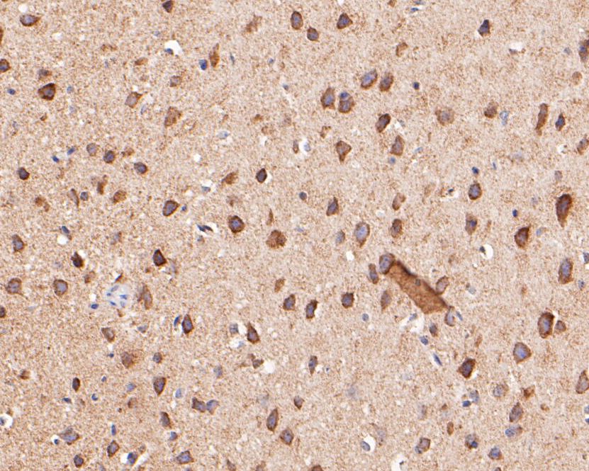
Fig6: Immunohistochemical analysis of paraffin-embedded mouse brain tissue using anti-KCNK18 antibody. The section was pre-treated using heat mediated antigen retrieval with Tris-EDTA buffer (pH 8.0-8.4) for 20 minutes.The tissues were blocked in 5% BSA for 30 minutes at room temperature, washed with ddH2O and PBS, and then probed with the primary antibody ( 1/200) for 30 minutes at room temperature. The detection was performed using an HRP conjugated compact polymer system. DAB was used as the chromogen. Tissues were counterstained with hematoxylin and mounted with DPX.
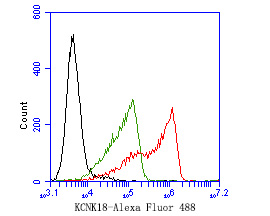
Fig7: Flow cytometric analysis of KCNK18 was done on SH-SY5Y cells. The cells were fixed, permeabilized and stained with the primary antibody ( 1/50) (red). After incubation of the primary antibody at room temperature for an hour, the cells were
- Background
-
References
- Czirjak G. et. al. The two-pore domain K+ channel, TRESK, is activated by the cytoplasmic calcium signal through calcineurin. J. Biol. Chem. 279:18550-18558(2004).
- Keshavaprasad B. et. al. Species-specific differences in response to anesthetics and other modulators by the K2P channel TRESK. Anesth. Analg. 101:1042-1049(2005).
- Bautista D.M. et. al. Pungent agents from Szechuan peppers excite sensory neurons by inhibiting two-pore potassium channels. Nat. Neurosci. 11:772-779(2008).
Related Products / Services
Please note: All products are "FOR RESEARCH USE ONLY AND ARE NOT INTENDED FOR DIAGNOSTIC OR THERAPEUTIC USE"