

-
Product Name
Anti-IL7 antibody
- Documents
-
Description
Mouse monoclonal antibody to IL7
-
Tested applications
ELISA, WB, ICC, IHC-P
-
Species reactivity
Human, Rat
-
Alternative names
IL-7 antibody
-
Isotype
IgG1
-
Preparation
This antigen of this antibody was recombinant protein
-
Clonality
Monoclonal
-
Formulation
Liquid, 1*TBS (pH7.4), 1%BSA, 40%Glycerol. Preservative: 0.17% Sodium Azide.
-
Storage instructions
Store at -20 ℃. Stable for 12 months from date of receipt.
-
Applications
WB: 1:500-1:1,000
ICC: 1:100-1:500
IHC-P: 1:50-1:200
ELISA: 1:5,000-1:10,000
-
Validations
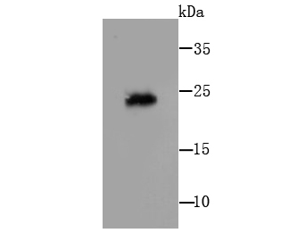
Fig1: Western blot analysis of IL7 on human thymus cell lysates using anti- IL7 antibody at 1/500 dilution.
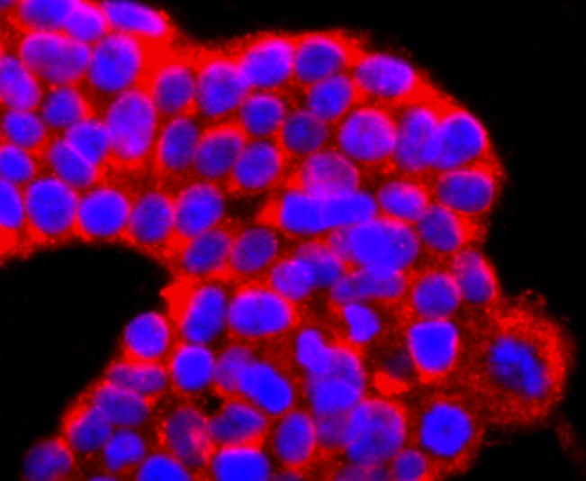
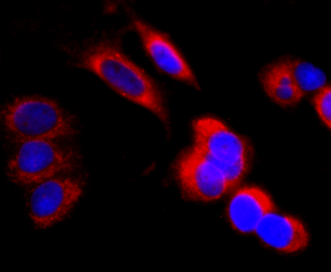
Fig2: ICC staining IL7 in 293T cells (red). The nuclear counter stain is DAPI (blue). Cells were fixed in paraformaldehyde, permeabilised with 0.25% Triton X100/PBS.
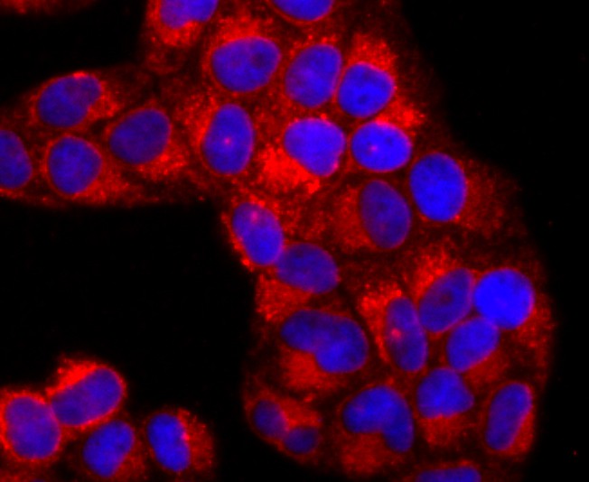
Fig3: ICC staining IL7 in Hela cells (red). The nuclear counter stain is DAPI (blue). Cells were fixed in paraformaldehyde, permeabilised with 0.25% Triton X100/PBS.
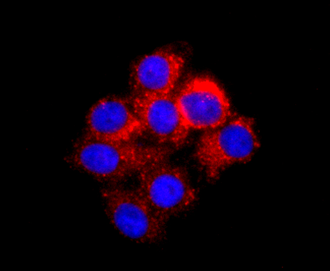
Fig4: ICC staining IL7 in HepG2 cells (red). The nuclear counter stain is DAPI (blue). Cells were fixed in paraformaldehyde, permeabilised with 0.25% Triton X100/PBS.
Fig5: ICC staining IL7 in MCF-7 cells (red). The nuclear counter stain is DAPI (blue). Cells were fixed in paraformaldehyde, permeabilised with 0.25% Triton X100/PBS.
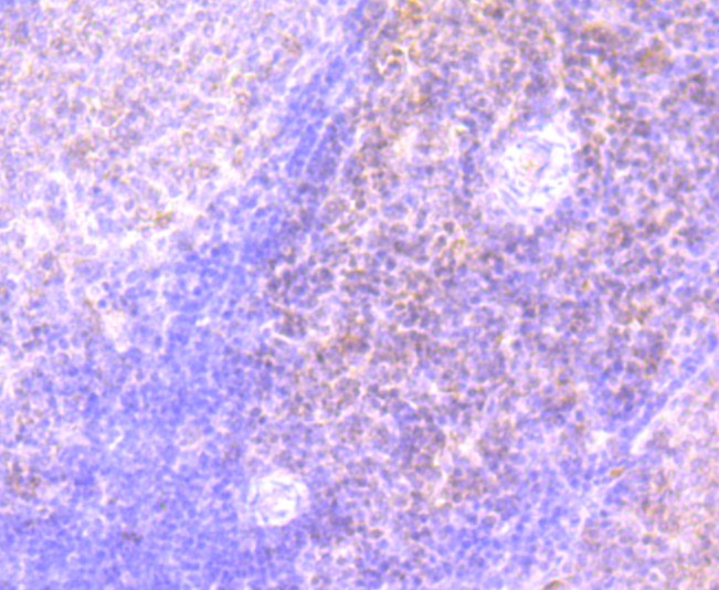
Fig6: Immunohistochemical analysis of paraffin-embedded rat spleen tissue using anti-IL7 antibody. Counter stained with hematoxylin.
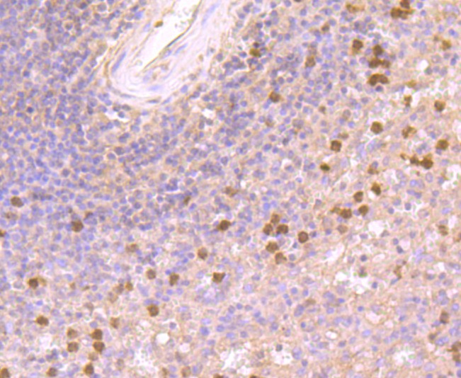
Fig7: Immunohistochemical analysis of paraffin-embedded human colon cancer tissue using anti-IL7 antibody. Counter stained with hematoxylin.
Fig8: Immunohistochemical analysis of paraffin-embedded human spleen tissue using anti-IL7 antibody. Counter stained with hematoxylin.
- Background
-
References
- Ming, J. et al. 2011. Interleukin-7 up-regulates cyclin D1 via activator protein-1 to promote proliferation of cell in lung cancer. Cancer Immunol Immunother.
- Zhang, Y. et al. 2015. Mutual enhancement of IL-2 and IL-7 on DNA vaccine immunogenicity mainly involves regulations on their receptor expression and receptor-expressing lymphocyte generation. Vaccine. 33: 3480-7.
Related Products / Services
Please note: All products are "FOR RESEARCH USE ONLY AND ARE NOT INTENDED FOR DIAGNOSTIC OR THERAPEUTIC USE"