
-
Product Name
Anti-IL6ST antibody
- Documents
-
Description
Mouse monoclonal antibody to IL6ST
-
Tested applications
WB, IHC-P, ICC
-
Species reactivity
Human
-
Alternative names
CD130 antibody; GP130 antibody; HIES4 antibody; CDW130 antibody; IL-6RB antibody; sGP130 antibody
-
Isotype
Mouse IgG2b
-
Preparation
This antigen of this antibody was recombinant protein
-
Clonality
Monoclonal
-
Formulation
Liquid, 1*PBS (pH7.4), 0.2% BSA, 50% Glycerol. Preservative: 0.05% Sodium Azide.
-
Storage instructions
Store at +4℃ after thawing. Aliquot store at -20℃ or -80℃. Avoid repeated freeze / thaw cycles.
-
Applications
WB: 1:500-1:2,000
ICC: 1:50-1:200
IHC-P: 1:50-1:200
-
Validations
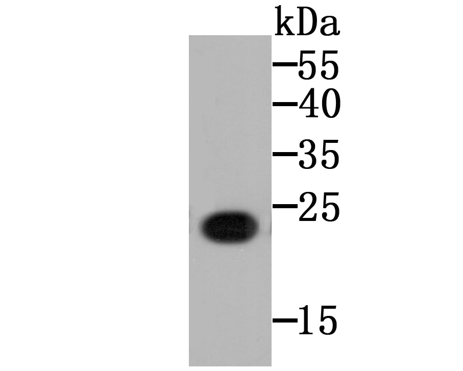
Fig1: Western blot analysis of CD130 on CD130 recombinant protein using anti-CD130 antibody at 1/5,000 dilution.
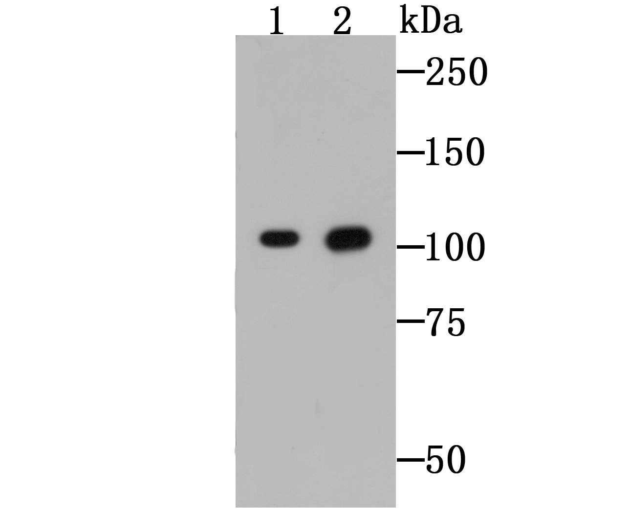
Fig2: Western blot analysis of CD130 on Raji cell (1) and mouse lung tissue (2) lysates using anti-CD130 antibody at 1/500 dilution.
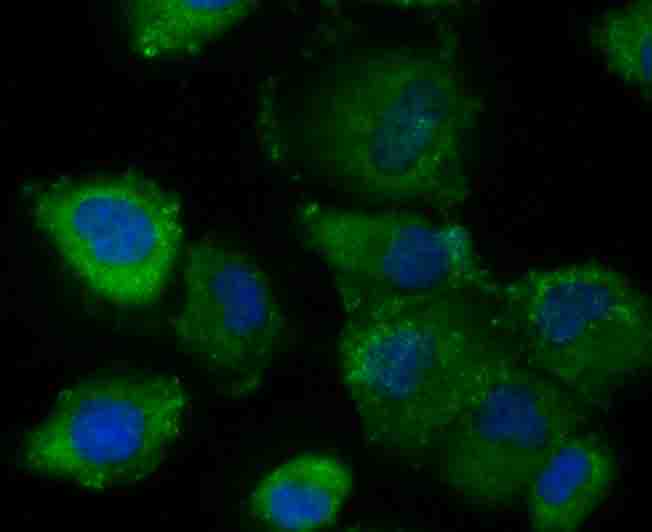
Fig3: ICC staining CD130 (green) in HUVEC cells. The nuclear counter stain is DAPI (blue). Cells were fixed in paraformaldehyde, permeabilised with 0.25% Triton X100/PBS.
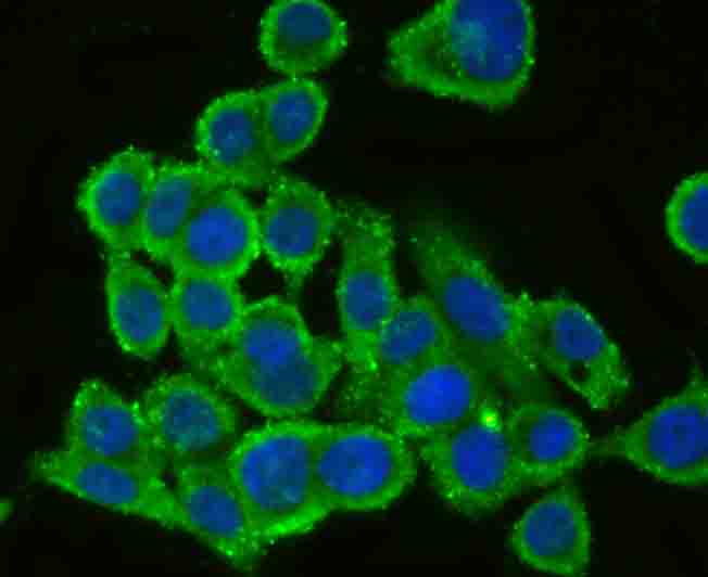
Fig4: ICC staining CD130 (green) in LOVO cells. The nuclear counter stain is DAPI (blue). Cells were fixed in paraformaldehyde, permeabilised with 0.25% Triton X100/PBS.
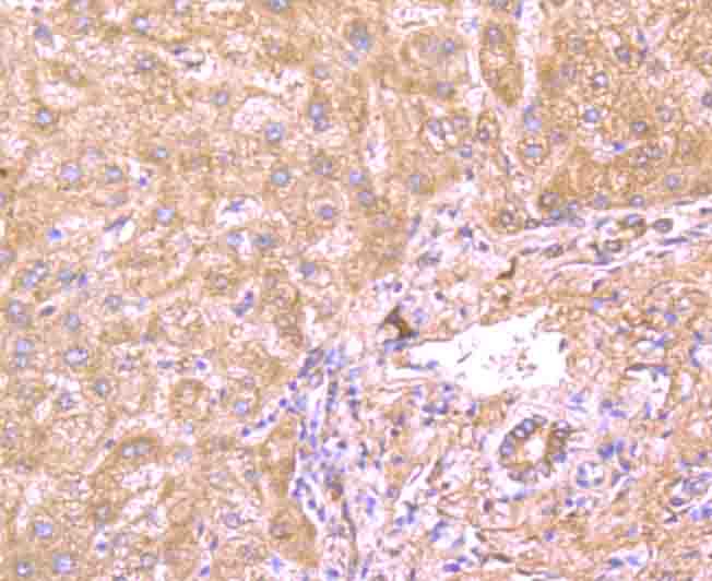
Fig5: Immunohistochemical analysis of paraffin-embedded human liver tissue using anti-CD130 antibody. Counter stained with hematoxylin.
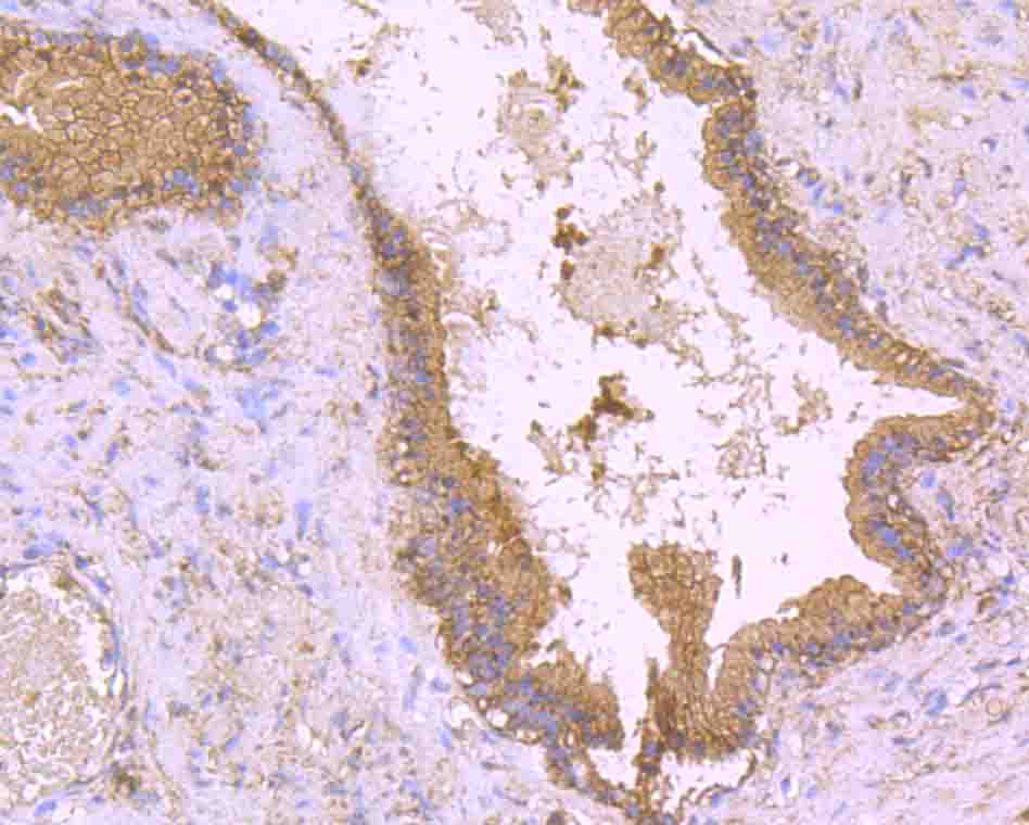
Fig6: Immunohistochemical analysis of paraffin-embedded human prostate tissue using anti-CD130 antibody. Counter stained with hematoxylin.
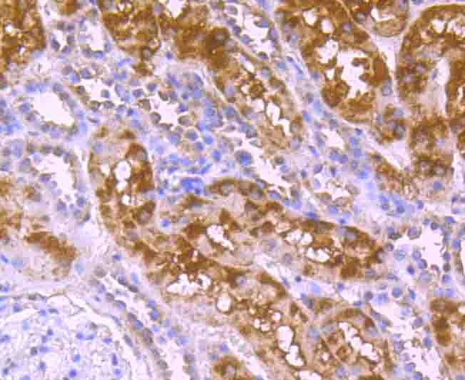
Fig7: Immunohistochemical analysis of paraffin-embedded human kidney tissue using anti-CD130 antibody. Counter stained with hematoxylin.
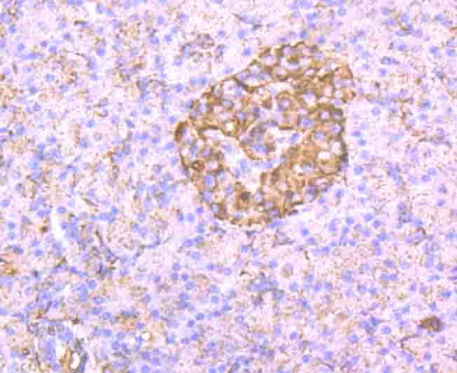
Fig8: Immunohistochemical analysis of paraffin-embedded human pancreas tissue using anti-CD130 antibody. Counter stained with hematoxylin.
- Background
-
References
- Schutt A et al. gp130 activation is regulated by D2-D3 interdomain connectivity. Biochem. J. 450:487-496 (2013).
- Waetzig GH et al. N-linked glycosylation is essential for the stability but not the signaling function of the interleukin-6 signal transducer glycoprotein 130. J. Biol. Chem. 285:1781-1789 (2010).
Related Products / Services
Please note: All products are "FOR RESEARCH USE ONLY AND ARE NOT INTENDED FOR DIAGNOSTIC OR THERAPEUTIC USE"