
-
Product Name
Anti-IL22 antibody
- Documents
-
Description
Rabbit polyclonal antibody to IL22
-
Tested applications
ELISA, WB, IHC-P
-
Species reactivity
Human, Mouse, Rat
-
Alternative names
TIFa antibody; IL-21 antibody; IL-22 antibody; ILTIF antibody; IL-TIF antibody; IL-D110 antibody; zcyto18 antibody; TIFIL-23 antibody
-
Isotype
Rabbit IgG
-
Preparation
This antigen of this antibody was recombinant protein within human il-22 aa 34-179.
-
Clonality
Polyclonal
-
Formulation
Liquid, 1*PBS (pH7.4), 0.2% BSA, 50% Glycerol. Preservative: 0.05% Sodium Azide.
-
Storage instructions
Store at +4℃ after thawing. Aliquot store at -20℃. Avoid repeated freeze / thaw cycles.
-
Applications
ELISA:1:5000-1:10,000
WB:1:500
IHC-P:1:50-1:200
-
Validations
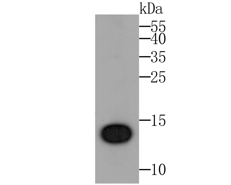
Fig1: Western blot analysis of IL-22 on IL22 recombinant protein. Proteins were transferred to a PVDF membrane and blocked with 5% BSA in PBS for 1 hour at room temperature. The primary antibody was used at a 1:500 dilution in 5% BSA at room temperature for 2 hours. Goat Anti-Rabbit IgG - HRP Secondary Antibody (HA1001) at 1:5,000 dilution was used for 1 hour at room temperature.
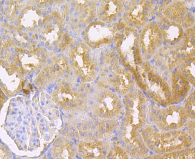
Fig2: Immunohistochemical analysis of paraffin-embedded rat kidney tissue using anti-IL-22 antibody. The section was pre-treated using heat mediated antigen retrieval with sodium citrate buffer (pH 6.0) for 20 minutes. The tissues were blocked in 5% BSA for 30 minutes at room temperature, washed with ddH2O and PBS, and then probed with the antibody at 1/200 dilution, for 30 minutes at room temperature and detected using an HRP conjugated compact polymer system. DAB was used as the chrogen. Counter stained with hematoxylin and mounted with DPX.
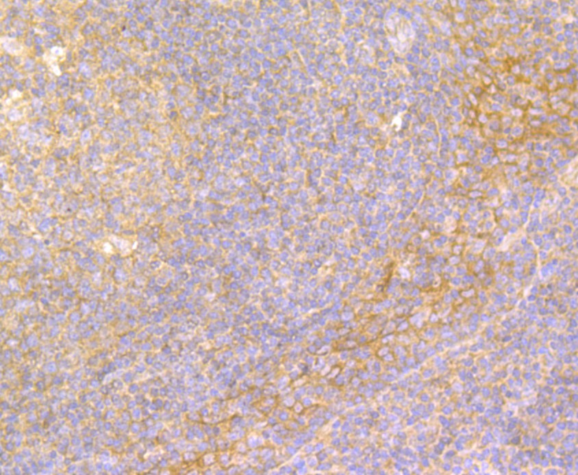
Fig3: Immunohistochemical analysis of paraffin-embedded human tonsil tissue using anti-IL-22 antibody. The section was pre-treated using heat mediated antigen retrieval with sodium citrate buffer (pH 6.0) for 20 minutes. The tissues were blocked in 5% BSA for 30 minutes at room temperature, washed with ddH2O and PBS, and then probed with the antibody at 1/200 dilution, for 30 minutes at room temperature and detected using an HRP conjugated compact polymer system. DAB was used as the chrogen. Counter stained with hematoxylin and mounted with DPX.
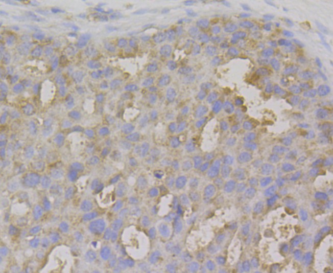
Fig4: Immunohistochemical analysis of paraffin-embedded human breasr cancer tissue using anti-IL-22 antibody. The section was pre-treated using heat mediated antigen retrieval with sodium citrate buffer (pH 6.0) for 20 minutes. The tissues were blocked in 5% BSA for 30 minutes at room temperature, washed with ddH2O and PBS, and then probed with the antibody at 1/200 dilution, for 30 minutes at room temperature and detected using an HRP conjugated compact polymer system. DAB was used as the chrogen. Counter stained with hematoxylin and mounted with DPX.
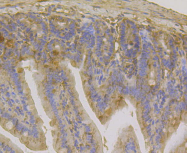
Fig5: Immunohistochemical analysis of paraffin-embedded mouse colon tissue using anti-IL-22 antibody. The section was pre-treated using heat mediated antigen retrieval with sodium citrate buffer (pH 6.0) for 20 minutes. The tissues were blocked in 5% BSA for 30 minutes at room temperature, washed with ddH2O and PBS, and then probed with the antibody at 1/200 dilution, for 30 minutes at room temperature and detected using an HRP conjugated compact polymer system. DAB was used as the chrogen. Counter stained with hematoxylin and mounted with DPX.
- Background
-
References
- Zhang Z et al. Signal peptide prediction based on analysis of experimentally verified cleavage sites. Protein Sci 13:2819-2824 (2004).
- Jones B C et al. Structure of IL-22 bound to its high-affinity IL-22R1 chain. Structure 16:1333-1344 (2008).
Related Products / Services
Please note: All products are "FOR RESEARCH USE ONLY AND ARE NOT INTENDED FOR DIAGNOSTIC OR THERAPEUTIC USE"