
-
Product Name
Anti-IGKC antibody
- Documents
-
Description
Rabbit monoclonal antibody to IGKC
-
Tested applications
WB, IHC-P
-
Species reactivity
Human
-
Alternative names
Km antibody; HCAK1 antibody; IGKCD antibody
-
Isotype
Rabbit IgG
-
Preparation
This antigen of this antibody was full length native human igg.
-
Clonality
Monoclonal
-
Formulation
Liquid, 1*TBS (pH7.4), 0.05% BSA, 40% Glycerol. Preservative: 0.05% Sodium Azide.
-
Storage instructions
Store at +4℃ after thawing. Aliquot store at -20℃. Avoid repeated freeze / thaw cycles.
-
Applications
WB: 1:500-1:2,000
IHC-P: 1:50-1:200
-
Validations
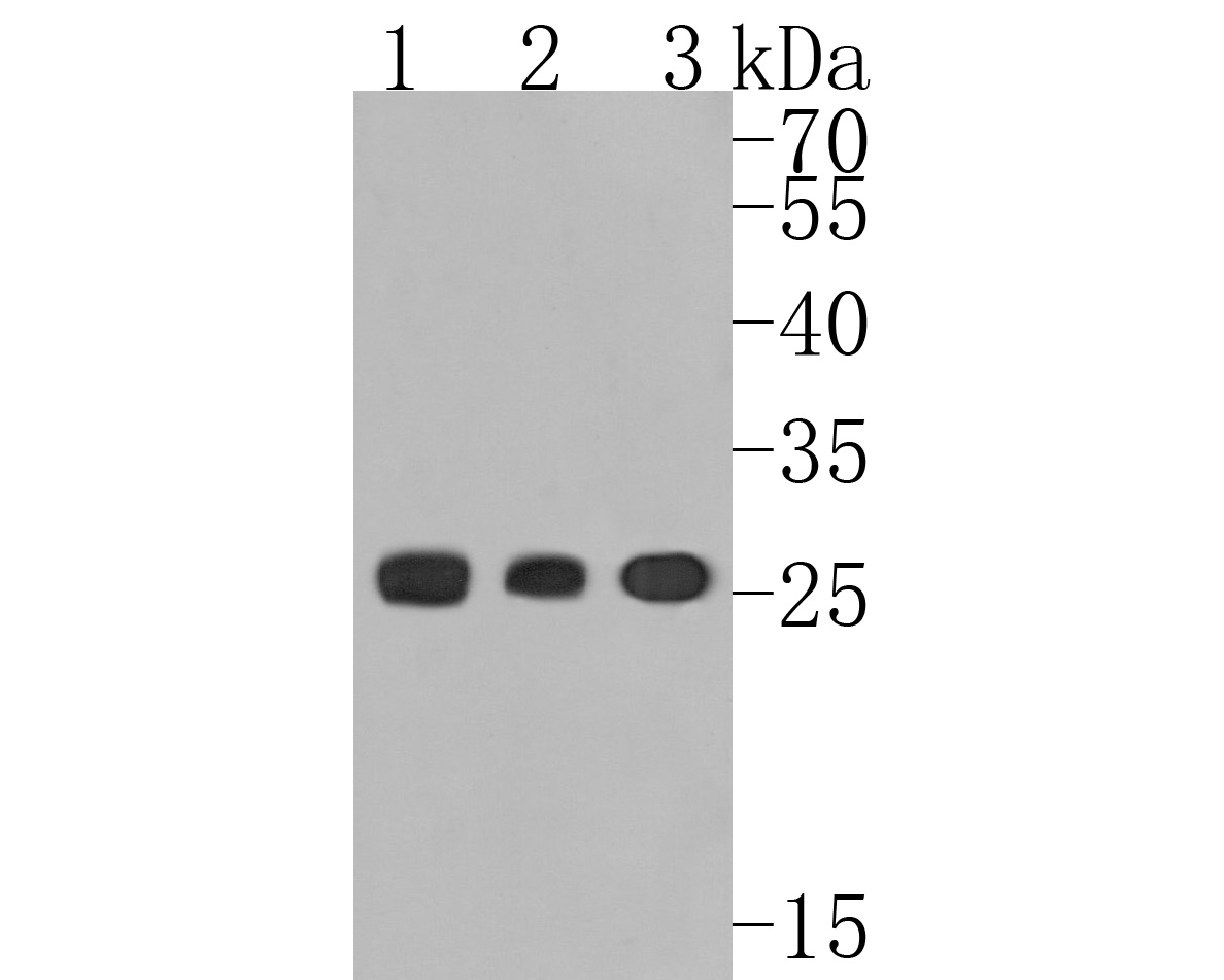
Fig1:; Western blot analysis of Human Kappa light chain on different lysates. Proteins were transferred to a PVDF membrane and blocked with 5% BSA in PBS for 1 hour at room temperature. The primary antibody ( 1/500) was used in 5% BSA at room temperature for 2 hours. Goat Anti-Rabbit IgG - HRP Secondary Antibody (HA1001) at 1:5,000 dilution was used for 1 hour at room temperature.; Positive control:; Lane 1: Human spleen tissue lysate; Lane 2: Human thymus tissue lysate; Lane 3: Human plasma tissue lysate
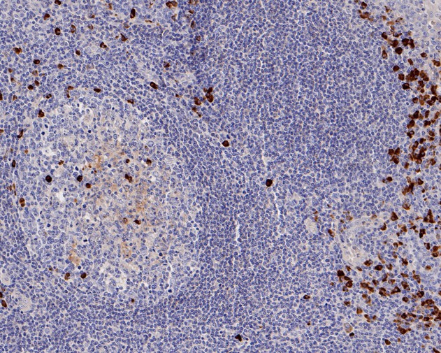
Fig2:; Immunohistochemical analysis of paraffin-embedded human tonsil tissue using anti-Human Kappa light chain antibody. The section was pre-treated using heat mediated antigen retrieval with Tris-EDTA buffer (pH 8.0-8.4) for 20 minutes.The tissues were blocked in 5% BSA for 30 minutes at room temperature, washed with ddH; 2; O and PBS, and then probed with the primary antibody ( 1/50) for 30 minutes at room temperature. The detection was performed using an HRP conjugated compact polymer system. DAB was used as the chromogen. Tissues were counterstained with hematoxylin and mounted with DPX.
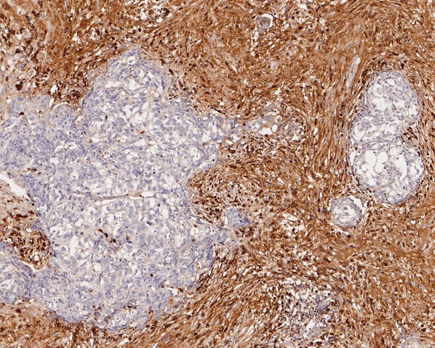
Fig3:; Immunohistochemical analysis of paraffin-embedded human breast carcinoma tissue using anti-Human Kappa light chain antibody. The section was pre-treated using heat mediated antigen retrieval with Tris-EDTA buffer (pH 8.0-8.4) for 20 minutes.The tissues were blocked in 5% BSA for 30 minutes at room temperature, washed with ddH; 2; O and PBS, and then probed with the primary antibody ( 1/50) for 30 minutes at room temperature. The detection was performed using an HRP conjugated compact polymer system. DAB was used as the chromogen. Tissues were counterstained with hematoxylin and mounted with DPX.
- Background
-
References
- Ma X. et. al. A novel xenograft mouse model for testing approaches targeting human kappa light-chain diseases. Gene Ther. 2019 May
- van der Kant R. et. al. Adaption of human antibody λ and κ light chain architectures to CDR repertoires. Protein Eng Des Sel. 2019 Dec
Related Products / Services
Please note: All products are "FOR RESEARCH USE ONLY AND ARE NOT INTENDED FOR DIAGNOSTIC OR THERAPEUTIC USE"