
-
Product Name
Anti-IGHM antibody
- Documents
-
Description
Rabbit monoclonal antibody to IGHM
-
Tested applications
WB, IHC-P, IP
-
Species reactivity
Human
-
Alternative names
MU antibody; VH antibody; AGM1 antibody
-
Isotype
Rabbit IgG
-
Preparation
This antigen of this antibody was recombinant protein within human igm from human serum (sigma#i8260).
-
Clonality
Monoclonal
-
Formulation
Liquid, 1*TBS (pH7.4), 0.05% BSA, 40% Glycerol. Preservative: 0.05% Sodium Azide.
-
Storage instructions
Store at +4℃ after thawing. Aliquot store at -20℃ or -80℃. Avoid repeated freeze / thaw cycles.
-
Applications
WB: 1:1,000-1:5,000
IHC-P: 1:50-1:200
-
Validations
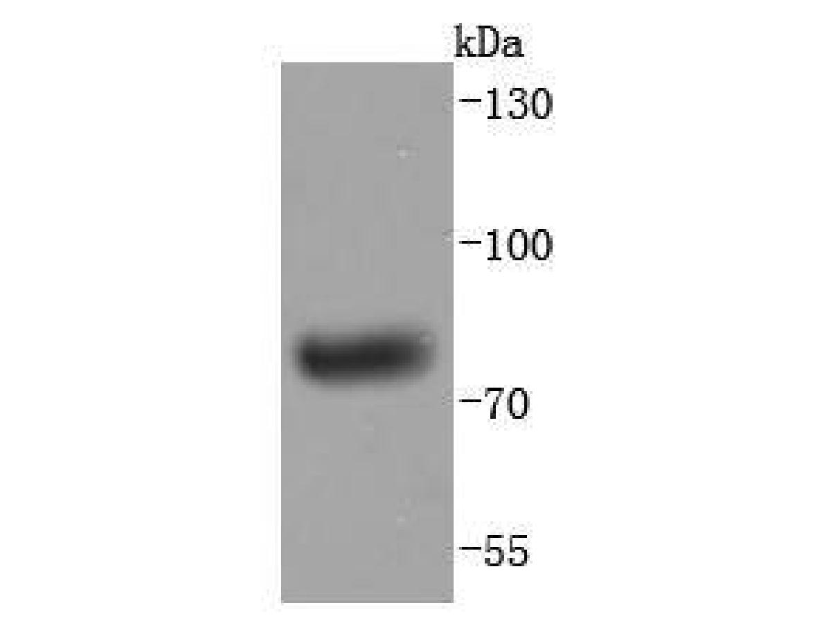
Fig1:; Western blot analysis of Human IgM on human plasma lysates. Proteins were transferred to a PVDF membrane and blocked with 5% BSA in PBS for 1 hour at room temperature. The primary antibody ( 1/500) was used in 5% BSA at room temperature for 2 hours. Goat Anti-Rabbit IgG - HRP Secondary Antibody (HA1001) at 1:200,000 dilution was used for 1 hour at room temperature.
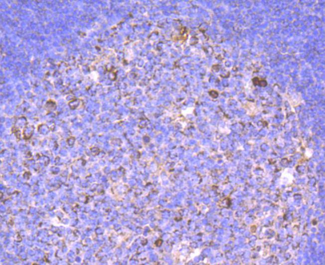
Fig2:; Immunohistochemical analysis of paraffin-embedded human tonsil tissue using anti-Human IgM antibody. The section was pre-treated using heat mediated antigen retrieval with Tris-EDTA buffer (pH 9.0) for 20 minutes.The tissues were blocked in 1% BSA for 30 minutes at room temperature, washed with ddH; 2; O and PBS, and then probed with the primary antibody ( 1/50) for 30 minutes at room temperature. The detection was performed using an HRP conjugated compact polymer system. DAB was used as the chromogen. Tissues were counterstained with hematoxylin and mounted with DPX.
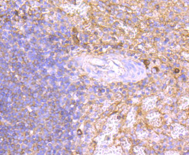
Fig3:; Immunohistochemical analysis of paraffin-embedded human spleen tissue using anti-Human IgM antibody. The section was pre-treated using heat mediated antigen retrieval with Tris-EDTA buffer (pH 9.0) for 20 minutes.The tissues were blocked in 1% BSA for 30 minutes at room temperature, washed with ddH; 2; O and PBS, and then probed with the primary antibody ( 1/50) for 30 minutes at room temperature. The detection was performed using an HRP conjugated compact polymer system. DAB was used as the chromogen. Tissues were counterstained with hematoxylin and mounted with DPX.
- Background
-
References
- Klasener K., et al. 2014. B cell activation involves nanoscale receptor reorganizations and inside-out signaling by Syk. Elife 3:e02069-e02069.
- Huang J., et al. 2014. Rearrangement and expression of the immunoglobulin mu-chain gene in human myeloid cells. Cell. Mol. Immunol. 11:94-104.
Related Products / Services
Please note: All products are "FOR RESEARCH USE ONLY AND ARE NOT INTENDED FOR DIAGNOSTIC OR THERAPEUTIC USE"