

-
Product Name
Anti-HECTD4 antibody
- Documents
-
Description
Mouse monoclonal antibody to HECTD4
-
Tested applications
WB, IHC-P, ICC, FC
-
Species reactivity
Human, Mouse
-
Alternative names
HEEL antibody; POTAGE antibody; C12ord51 antibody; C12orf51 antibody
-
Isotype
Mouse IgG1
-
Preparation
This antigen of this antibody was recombinant protein
-
Clonality
Monoclonal
-
Formulation
Liquid, 1*PBS (pH7.4), 0.2% BSA, 50% Glycerol. Preservative: 0.05% Sodium Azide.
-
Storage instructions
Store at +4℃ after thawing. Aliquot store at -20℃ or -80℃. Avoid repeated freeze / thaw cycles.
-
Applications
WB: 1:500
ICC: 1:50
IHC-P: 1:50-1:100
FC: 1:50-1:100
-
Validations
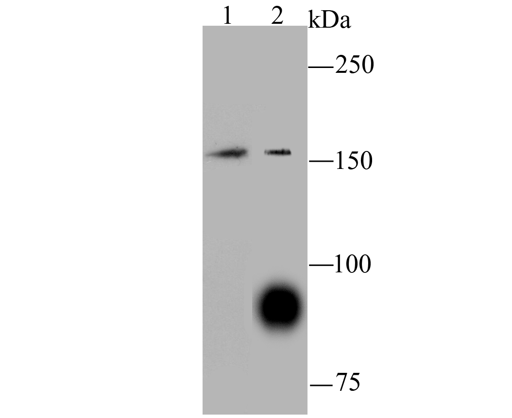
Fig1: Western blot analysis of C12orf51 on SH-SY5Y (1) and A549 (2) using anti-C12orf51 antibody at 1/100 dilution.
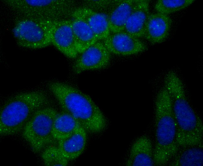
Fig2: ICC staining C12orf51 (green) in HepG2 cells. The nuclear counter stain is DAPI (blue). Cells were fixed in paraformaldehyde, permeabilised with 0.25% Triton X100/PBS.
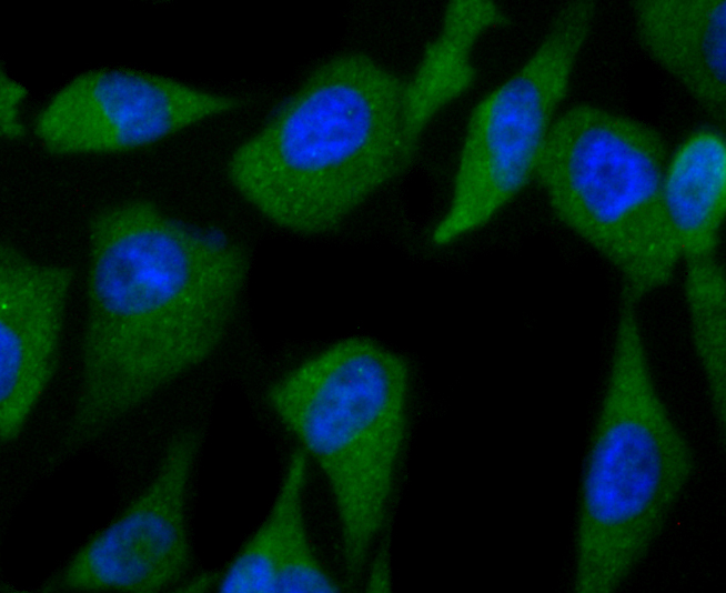
Fig3: ICC staining C12orf51 (green) in PC-3M cells. The nuclear counter stain is DAPI (blue). Cells were fixed in paraformaldehyde, permeabilised with 0.25% Triton X100/PBS.
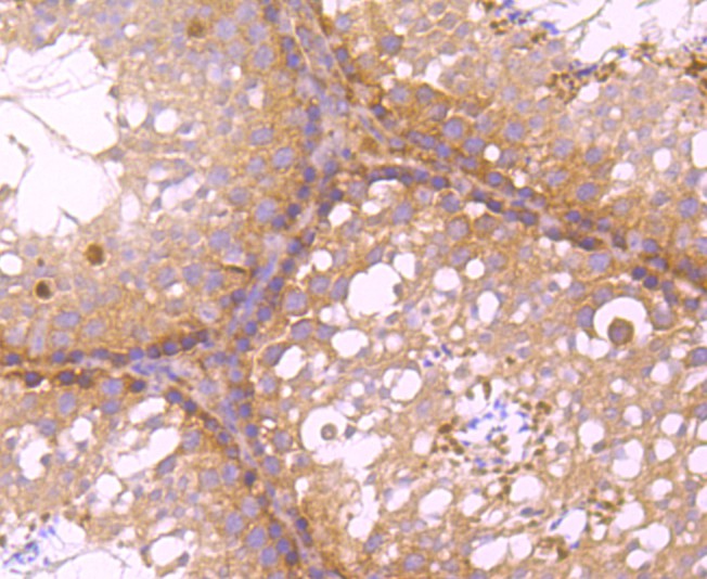
Fig4: Immunohistochemical analysis of paraffin-embedded human colon cancer tissue using anti-C12orf51 antibody. Counter stained with hematoxylin.
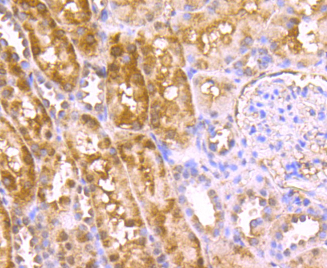
Fig5: Immunohistochemical analysis of paraffin-embedded human kidney tissue using anti-C12orf51 antibody. Counter stained with hematoxylin.
Fig6: Immunohistochemical analysis of paraffin-embedded mouse testis tissue using anti-C12orf51 antibody. Counter stained with hematoxylin.
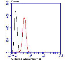
Fig7: Flow cytometric analysis of SH-SY5Y cells with C12orf51 antibody at 1/100 dilution (red) compared with an unlabelled control (cells without incubation with primary antibody; black).
- Background
-
References
- Eom SY et al.Exome-wide association study identifies genetic polymorphisms of C12orf51, MYL2, and ALDH2 associated with blood lead levels in the general Korean population. Environ Health 16(1):11 (2017).
- Kim J et al.No Interaction with Alcohol Consumption, but Independent Effect of C12orf51 (HECTD4) on Type 2 Diabetes Mellitus in Korean Adults Aged 40-69 Years: The KoGES_Ansan and Ansung Study. PLoS One 11(2):e0149321 (2016).
Related Products / Services
Please note: All products are "FOR RESEARCH USE ONLY AND ARE NOT INTENDED FOR DIAGNOSTIC OR THERAPEUTIC USE"