
-
Product Name
Anti-H1-4 antibody
- Documents
-
Description
Rabbit monoclonal antibody to H1-4
-
Tested applications
WB, ICC/IF, IHC-P
-
Species reactivity
Human, Mouse, Rat
-
Alternative names
H1E antibody; H1.4 antibody; H1F4 antibody; RMNS antibody; H1s-4 antibody; HIST1H1E antibody; dJ221C16.5 antibody
-
Isotype
Rabbit IgG
-
Preparation
This antigen of this antibody was synthetic phospho-peptide corresponding to residues surrounding thr17 of human h1.4.
-
Clonality
Monoclonal
-
Formulation
Liquid, 1*TBS (pH7.4), 0.05% BSA, 40% Glycerol. Preservative: 0.05% Sodium Azide.
-
Storage instructions
Store at +4℃ after thawing. Aliquot store at -20℃ or -80℃. Avoid repeated freeze / thaw cycles.
-
Applications
WB: 1:500-1:1,000
ICC/IF: 1:50-1:200
IHC-P: 1:50-1:200
-
Validations
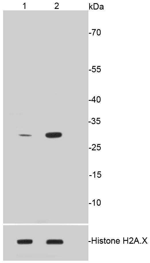
Fig1:; Western blot analysis of Phospho-Histone H1.3(T17)+Histone H1.4(T17) on different lysates. Proteins were transferred to a PVDF membrane and blocked with 5% BSA in PBS for 1 hour at room temperature. The primary antibody ( 1/500) was used in 5% BSA at room temperature for 2 hours. Goat Anti-Rabbit IgG - HRP Secondary Antibody (HA1001) at 1:40,000 dilution was used for 1 hour at room temperature.; Positive control:; Lane 1: Untreated CRC whole cell lysates; Lane 2: CRC cells treated with 1.5ug/ml Colcemid for 12 hours whole cell lysates
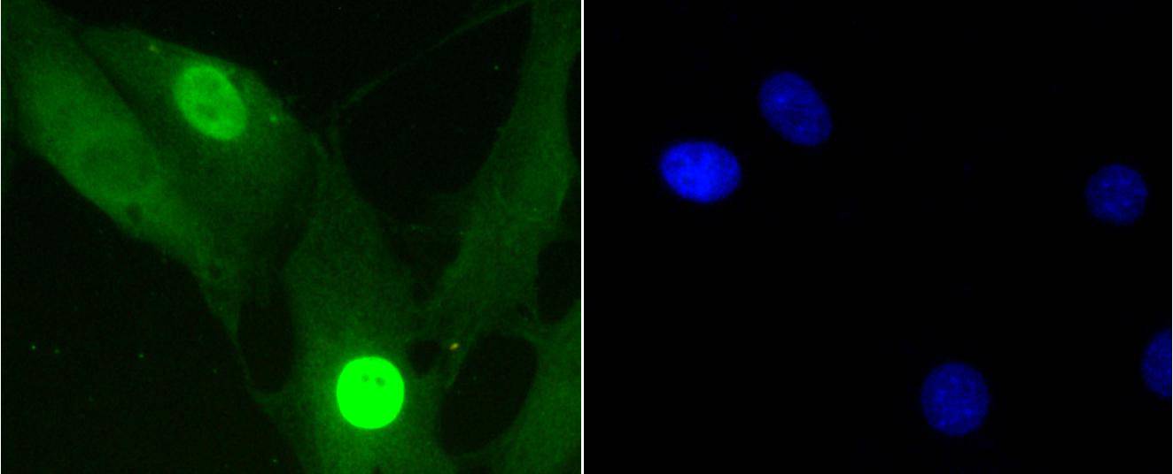
Fig2:; ICC staining of Phospho-Histone H1.3(T17)+Histone H1.4(T17) in NIH/3T3 cells (green). Formalin fixed cells were permeabilized with 0.1% Triton X-100 in TBS for 10 minutes at room temperature and blocked with 1% Blocker BSA for 15 minutes at room temperature. Cells were probed with the primary antibody ( 1/50) for 1 hour at room temperature, washed with PBS. Alexa Fluor®488 Goat anti-Rabbit IgG was used as the secondary antibody at 1/1,000 dilution. The nuclear counter stain is DAPI (blue).
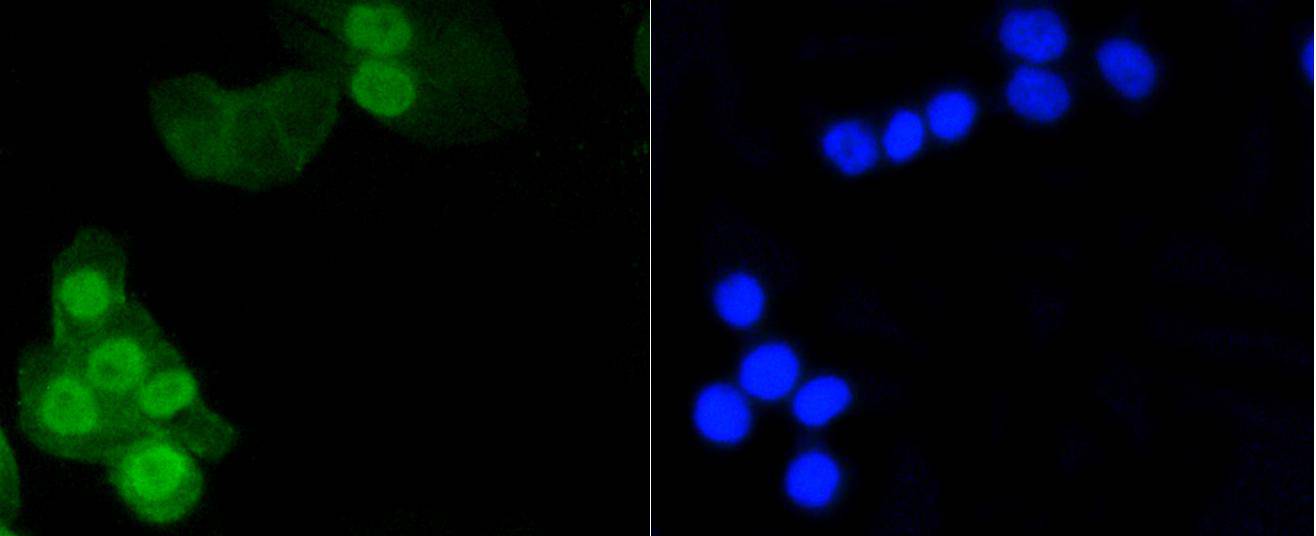
Fig3:; ICC staining of Phospho-Histone H1.3(T17)+Histone H1.4(T17) in CRC cells (green). Formalin fixed cells were permeabilized with 0.1% Triton X-100 in TBS for 10 minutes at room temperature and blocked with 1% Blocker BSA for 15 minutes at room temperature. Cells were probed with the primary antibody ( 1/50) for 1 hour at room temperature, washed with PBS. Alexa Fluor®488 Goat anti-Rabbit IgG was used as the secondary antibody at 1/1,000 dilution. The nuclear counter stain is DAPI (blue).
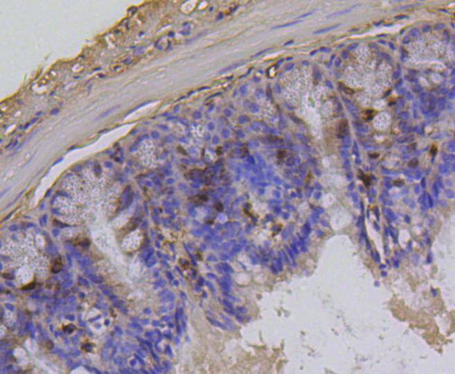
Fig4:; Immunohistochemical analysis of paraffin-embedded mouse colon cancer tissue using anti-Phospho-Histone H1.3(T17)+Histone H1.4(T17) antibody. Counter stained with hematoxylin.
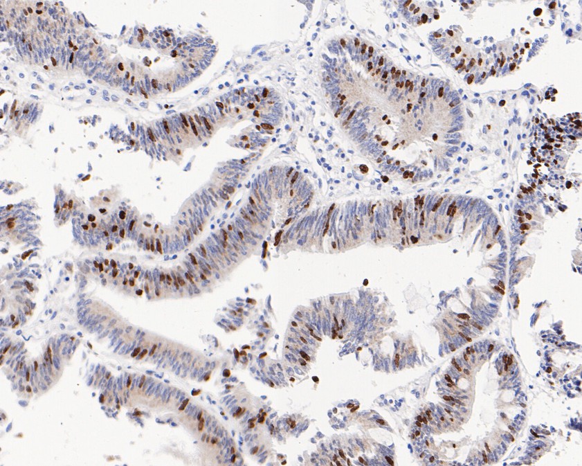
Fig5:; Immunohistochemical analysis of paraffin-embedded human colon carcinoma tissue using anti-Phospho-Histone H1.3(T17)+Histone H1.4(T17) antibody. The section was pre-treated using heat mediated antigen retrieval with sodium citrate buffer (pH 6.0) for 20 minutes. The tissues were blocked in 5% BSA for 30 minutes at room temperature, washed with ddH; 2; O and PBS, and then probed with the primary antibody ( 1/200) for 30 minutes at room temperature. The detection was performed using an HRP conjugated compact polymer system. DAB was used as the chromogen. Tissues were counterstained with hematoxylin and mounted with DPX.
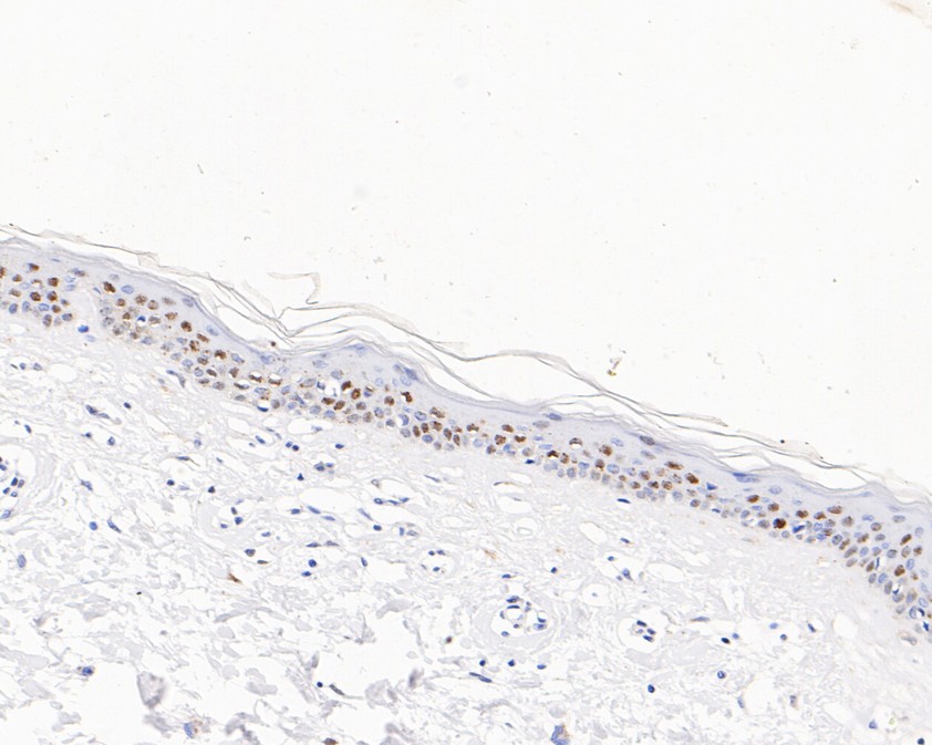
Fig6:; Immunohistochemical analysis of paraffin-embedded human skin tissue using anti-Phospho-Histone H1.3(T17)+Histone H1.4(T17) antibody. The section was pre-treated using heat mediated antigen retrieval with sodium citrate buffer (pH 6.0) for 20 minutes. The tissues were blocked in 5% BSA for 30 minutes at room temperature, washed with ddH; 2; O and PBS, and then probed with the primary antibody ( 1/200) for 30 minutes at room temperature. The detection was performed using an HRP conjugated compact polymer system. DAB was used as the chromogen. Tissues were counterstained with hematoxylin and mounted with DPX.
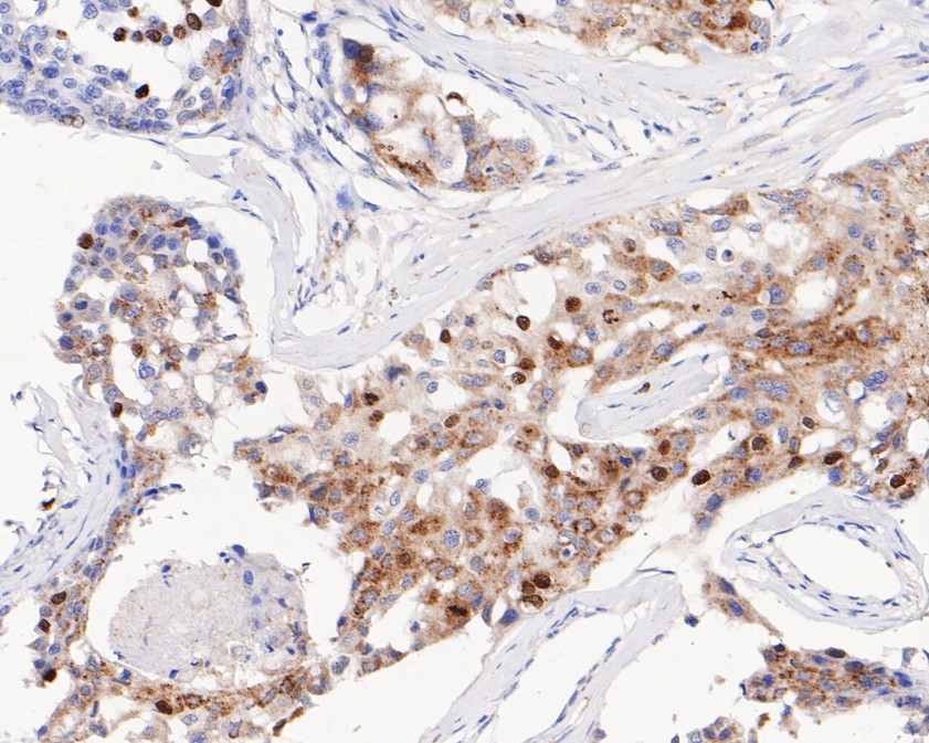
Fig7:; Immunohistochemical analysis of paraffin-embedded human breast carcinoma tissue using anti-Phospho-Histone H1.3(T17)+Histone H1.4(T17) antibody. The section was pre-treated using heat mediated antigen retrieval with sodium citrate buffer (pH 6.0) for 20 minutes. The tissues were blocked in 5% BSA for 30 minutes at room temperature, washed with ddH; 2; O and PBS, and then probed with the primary antibody ( 1/200) for 30 minutes at room temperature. The detection was performed using an HRP conjugated compact polymer system. DAB was used as the chromogen. Tissues were counterstained with hematoxylin and mounted with DPX.
- Background
-
References
- Vaca Jacome A.S., Rabilloud T., Schaeffer-Reiss C., et al. N-terminome analysis of the human mitochondrial proteome. Proteomics 15:2519-2524(2015).
- Bienvenut W.V., Sumpton D., Martinez A., et al. Comparative large-scale characterisation of plant vs. mammal proteins reveals similar and idiosyncratic N-alpha acetylation features. Mol. Cell. Proteomics 11:M111.015131-M111.015131(2012).
Related Products / Services
Please note: All products are "FOR RESEARCH USE ONLY AND ARE NOT INTENDED FOR DIAGNOSTIC OR THERAPEUTIC USE"