

-
Product Name
Anti-Gabbr2 antibody
- Documents
-
Description
Rabbit monoclonal antibody to Gabbr2
-
Tested applications
WB, IHC-P, FC, ICC
-
Species reactivity
Human, Mouse, Rat
-
Alternative names
Gb2 antibody; Gpr antibody; Gaba antibody; Gm425 antibody; Gpr51 antibody; GABABR2 antibody
-
Isotype
Rabbit IgG
-
Preparation
This antigen of this antibody was recombinant protein
-
Clonality
Monoclonal
-
Formulation
Liquid, 1*TBS (pH7.4), 0.05% BSA, 40% Glycerol. Preservative: 0.05% Sodium Azide.
-
Storage instructions
Store at +4℃ after thawing. Aliquot store at -20℃ or -80℃. Avoid repeated freeze / thaw cycles.
-
Applications
WB: 1:500-1:1,000
IHC-P: 1:50-1:200
FC: 1:50-1:100
ICC: 1:50-1:100
-
Validations
Fig1: Western blot analysis of GABA B Receptor 2 on mouse cerebellum tissue lysate using anti- GABA B Receptor 2 antibody at 1/500 dilution.
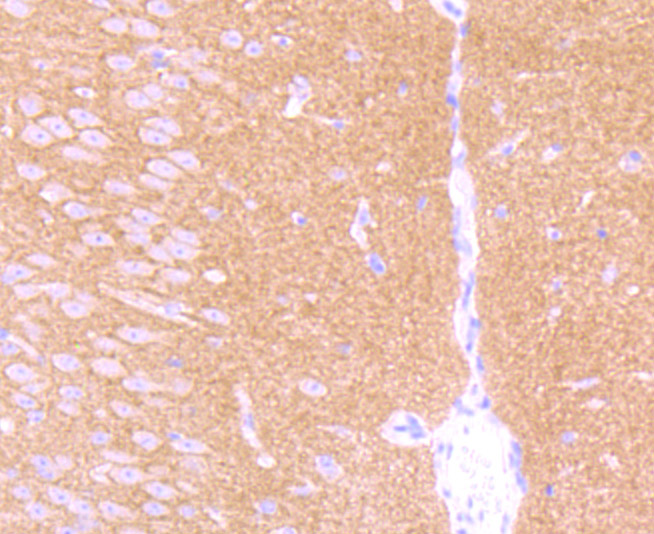
Fig2: Immunohistochemical analysis of paraffin-embedded mouse brain tissue using anti- GABA B Receptor 2 antibody. Counter stained with hematoxylin.
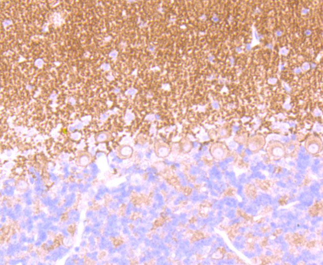
Fig3: Immunohistochemical analysis of paraffin-embedded mouse cerebellum tissue using anti- GABA B Receptor 2 antibody. Counter stained with hematoxylin.
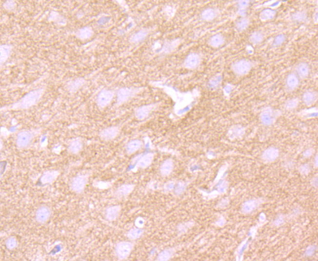
Fig4: Immunohistochemical analysis of paraffin-embedded rat brain tissue using anti- GABA B Receptor 2 antibody. Counter stained with hematoxylin.
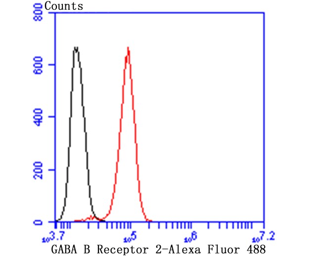
Fig5: Flow cytometric analysis of SH-SY-5Y cells with GABA B Receptor 2 antibody at 1/50 dilution (red) compared with an unlabelled control (cells without incubation with primary antibody; black). Alexa Fluor 488-conjugated goat anti rabbit IgG was used as the secondary antibody.
- Background
-
References
- White J H et al. Heterodimerization is required for the formation of a functional GABA(B) receptor. Nature 396:679-682 (1998).
- Martin S C et al. Molecular identification of the human GABABR2: cell surface expression and coupling to adenylyl cyclase in the absence of GABABR1. Mol Cell Neurosci 13:180-191 (1999).
Related Products / Services
Please note: All products are "FOR RESEARCH USE ONLY AND ARE NOT INTENDED FOR DIAGNOSTIC OR THERAPEUTIC USE"