
-
Product Name
Anti-FBXO5 antibody
- Documents
-
Description
Rabbit monoclonal antibody to FBXO5
-
Tested applications
WB, ICC, IHC-P
-
Species reactivity
Human, Mouse, Rat
-
Alternative names
EMI1 antibody; FBX5 antibody; Fbxo31 antibody
-
Isotype
Rabbit IgG
-
Preparation
This antigen of this antibody was recombinant protein within human emi1 aa 50-250.
-
Clonality
Monoclonal
-
Formulation
Liquid, 1*TBS (pH7.4), 0.05% BSA, 40% Glycerol. Preservative: 0.05% Sodium Azide.
-
Storage instructions
Store at +4℃ after thawing. Aliquot store at -20℃. Avoid repeated freeze / thaw cycles.
-
Applications
ICC: 1:50-1:200
IHC-P: 1:50-1:200
WB: 1:500
-
Validations
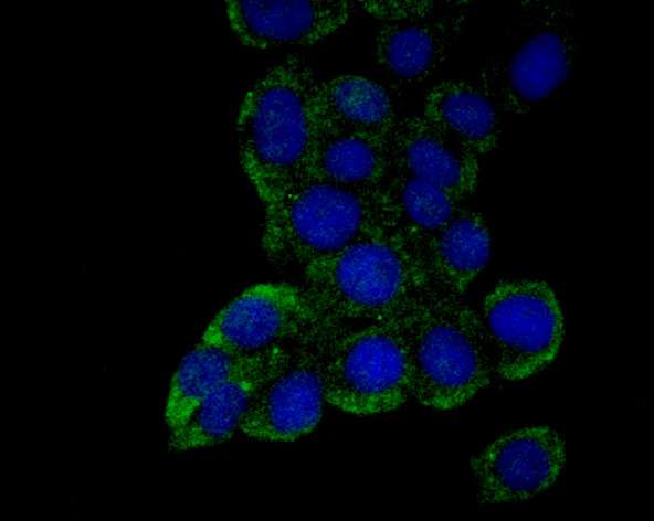
Fig1: ICC staining Emi1 in LOVO cells (green). The nuclear counter stain is DAPI (blue). Cells were fixed in paraformaldehyde, permeabilised with 0.25% Triton X100/PBS.
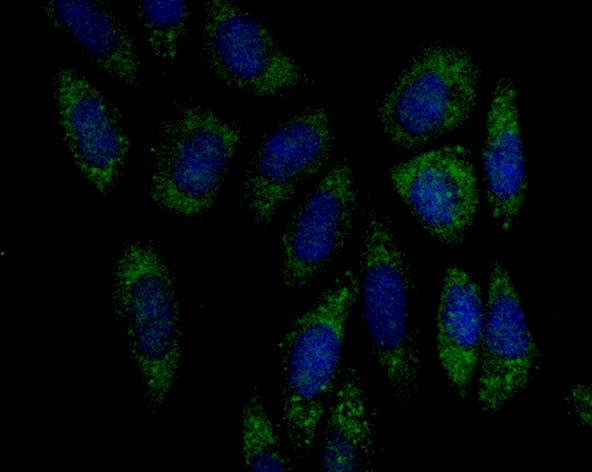
Fig2: ICC staining Emi1 in Siha cells (green). The nuclear counter stain is DAPI (blue). Cells were fixed in paraformaldehyde, permeabilised with 0.25% Triton X100/PBS.
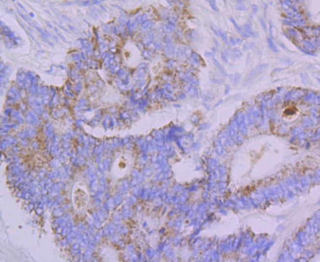
Fig3: Immunohistochemical analysis of paraffin-embedded human colon cancer tissue using anti-Emi1 antibody. Counter stained with hematoxylin.
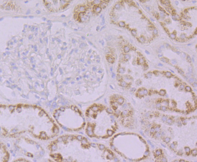
Fig4: Immunohistochemical analysis of paraffin-embedded human kidney tissue using anti-Emi1 antibody. Counter stained with hematoxylin.
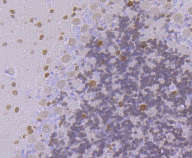
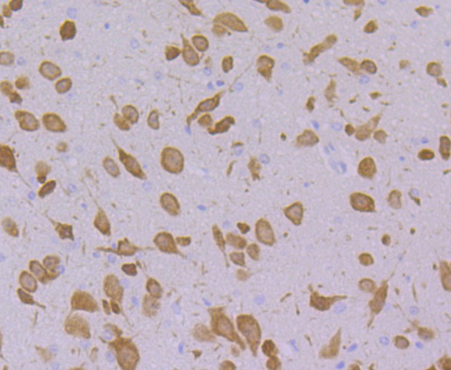
Fig5: Immunohistochemical analysis of paraffin-embedded rat cerebellum tissue using anti-Emi1 antibody. Counter stained with hematoxylin.
Fig6: Immunohistochemical analysis of paraffin-embedded mouse cerebellum tissue using anti-Emi1 antibody. Counter stained with hematoxylin.
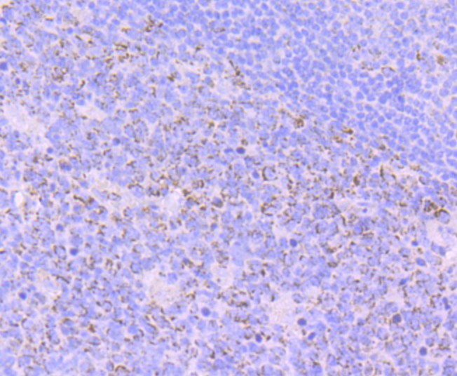
Fig7: Immunohistochemical analysis of paraffin-embedded human tonsil tissue using anti-Emi1 antibody. Counter stained with hematoxylin.
- Background
-
References
- Hsu J Y et al. E2F-dependent accumulation of hEmi1 regulates S phase entry by inhibiting APC(Cdh1). Nat Cell Biol 4:358-366 (2002).
- Miller J J et al. Emi1 stably binds and inhibits the anaphase-promoting complex/cyclosome as a pseudosubstrate inhibitor. Genes Dev 20:2410-2420 (2006).
Related Products / Services
Please note: All products are "FOR RESEARCH USE ONLY AND ARE NOT INTENDED FOR DIAGNOSTIC OR THERAPEUTIC USE"