
-
Product Name
Anti-ERAS antibody
- Documents
-
Description
Mouse monoclonal antibody to ERAS
-
Tested applications
ICC, IHC-P
-
Species reactivity
Human
-
Alternative names
HRAS2 antibody; HRASP antibody
-
Isotype
Mouse IgG1
-
Preparation
This antigen of this antibody was recombinant protein.
-
Clonality
Monoclonal
-
Formulation
Liquid, 1*PBS (pH7.4), 0.2% BSA, 40% Glycerol. Preservative: 0.05% Sodium Azide.
-
Storage instructions
Store at +4℃ after thawing. Aliquot store at -20℃. Avoid repeated freeze / thaw cycles.
-
Applications
ELISA:
WB:
IP:
ICC:1:200
IF:
IHC-P:1:200-1:500
FC:
-
Validations
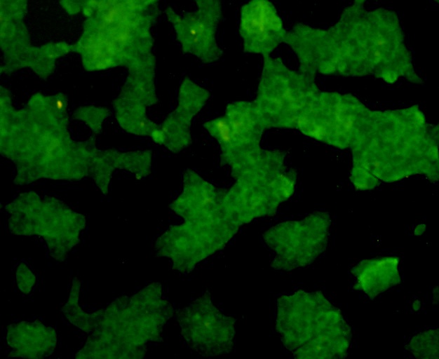
Fig1: ICC staining ERAS in NCCIT cells (green). Formalin fixed cells were permeabilized with 0.1% Triton X-100 in TBS for 10 minutes at room temperature and blocked with 1% Blocker BSA for 15 minutes at room temperature. Cells were probed with ERAS monoclonal antibody at a dilution of 1:200 for 1 hour at room temperature, washed with PBS. Alexa Fluorc™ 488 Goat anti-Mouse IgG was used as the secondary antibody at 1/100 dilution.
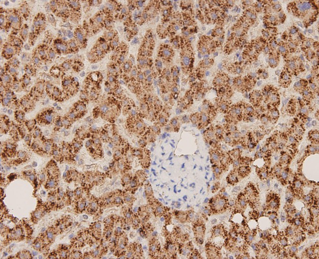
Fig2: Immunohistochemical analysis of paraffin-embedded human liver carcinoma tissue using anti-ERAS antibody. The section was pre-treated using heat mediated antigen retrieval with Tris-EDTA buffer (pH 8.0-8.4) for 20 minutes.The tissues were blocked in 5% BSA for 30 minutes at room temperature, washed with ddH2O and PBS, and then probed with the antibody at 1/100 dilution, for 30 minutes at room temperature and detected using an HRP conjugated compact polymer system. DAB was used as the chrogen. Counter stained with hematoxylin and mounted with DPX.
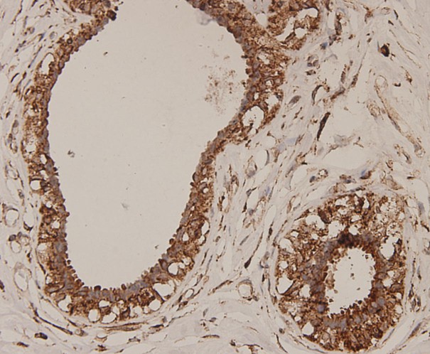
Fig3: Immunohistochemical analysis of paraffin-embedded human breast carcinoma tissue using anti-ERAS antibody. The section was pre-treated using heat mediated antigen retrieval with Tris-EDTA buffer (pH 8.0-8.4) for 20 minutes.The tissues were blocked in 5% BSA for 30 minutes at room temperature, washed with ddH2O and PBS, and then probed with the antibody at 1/100 dilution, for 30 minutes at room temperature and detected using an HRP conjugated compact polymer system. DAB was used as the chrogen. Counter stained with hematoxylin and mounted with DPX.
- Background
-
References
- Takahashi K et al. Role of Eras in promoting tumor-like properties in mouse embryonic stem cells. Nature 423:541-545 (2003).
- Liu Y et al. Role of the ERas gene in gastric cancer cells. Oncol Rep 30(1):50-6 (2013).
- Kubota E et al. Role of ES cell-expressed Ras (ERas) in tumorigenicity of gastric cancer. Am J Pathol 177(2):955-63 (2010).
Related Products / Services
Please note: All products are "FOR RESEARCH USE ONLY AND ARE NOT INTENDED FOR DIAGNOSTIC OR THERAPEUTIC USE"