
-
Product Name
Anti-Emp1 antibody
- Documents
-
Description
Rabbit polyclonal antibody to Emp1
-
Tested applications
WB, IHC-P, FC
-
Species reactivity
Human, Mouse, Rat
-
Alternative names
TMP antibody; I-8-09 antibody
-
Isotype
Rabbit IgG
-
Preparation
This antigen of this antibody was klh conjugated synthetic peptide derived from mouse emp1 101-160/160
-
Clonality
Polyclonal
-
Formulation
Liquid, 0.01M TBS(pH7.4) with 1% BSA, 0.03% Proclin300 and 50% Glycerol.
-
Storage instructions
Store at -20℃ for one year. Avoid repeated freeze/thaw cycles. The lyophilized antibody is stable at room temperature for at least one month and for greater than a year when kept at -20℃. When reconstituted in sterile pH 7.4 0.01M PBS or diluent of antibody the antibody is stable for at least two weeks at 2-4℃.
-
Applications
WB:1:500-2000
IHC-P:1:400-800
FC:3ug/Test
-
Validations
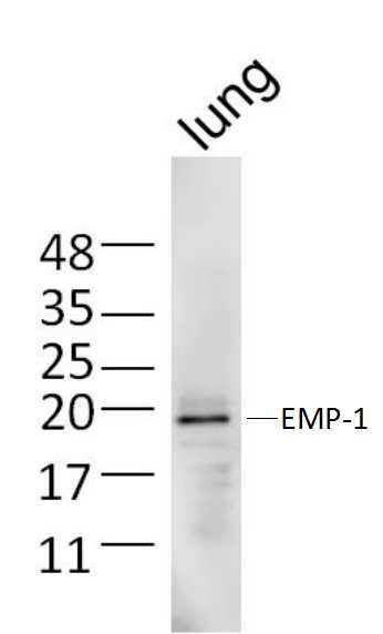
Fig1: Sample:; Lung (Mouse) Lysate at 40 ug; Primary: Anti-EMP-1 at 1/300 dilution; Secondary: IRDye800CW Goat Anti-Rabbit IgG at 1/20000 dilution; Predicted band size: 17 kD; Observed band size: 19 kD
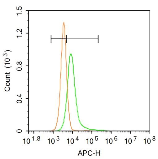
Fig2: Blank control: A431.; Primary Antibody (green line): Rabbit Anti-EMP-1 antibody ; Dilution: 3μg /10^6 cells;; Isotype Control Antibody (orange line): Rabbit IgG .; Secondary Antibody: Goat anti-rabbit IgG-AF647; Dilution: 3μg /test.; Protocol; The cells were incubated in 5%BSA to block non-specific protein-protein interactions for 30 min at at room temperature .Cells stained with Primary Antibody for 30 min at room temperature. The secondary antibody used for 40 min at room temperature. Acquisition of 20,000 events was performed.
- Background
-
References
- Chang Liu, Xiaojun Wei. The Prognostic Value of Epithelial Membrane Protein 1 (EMP-1) in Patients with Laryngeal Carcinoma. Med Sci Monit. 2017; 23: 3795–3800.
- Mohammad Khusni B. Ahmat Amin. Epithelial membrane protein 1 promotes tumor metastasis by enhancing cell migration via copine-III and Rac1. Oncogene. 2018; 37(40): 5416–5434.
Related Products / Services
Please note: All products are "FOR RESEARCH USE ONLY AND ARE NOT INTENDED FOR DIAGNOSTIC OR THERAPEUTIC USE"