
-
Product Name
Anti-CNTN2 antibody
- Documents
-
Description
Rabbit polyclonal antibody to CNTN2
-
Tested applications
WB, ICC
-
Species reactivity
Human, Mouse, Rat
-
Alternative names
AXT antibody; TAX antibody; TAX1 antibody; FAME5 antibody; TAG-1 antibody
-
Isotype
Rabbit IgG
-
Preparation
This antigen of this antibody was recombinant protein within human tag1 aa 40-230.
-
Clonality
Polyclonal
-
Formulation
Liquid, 1*TBS (pH7.4), 0.2% BSA, 50% Glycerol. Preservative: 0.05% Sodium Azide.
-
Storage instructions
Store at +4℃ after thawing. Aliquot store at -20℃. Avoid repeated freeze / thaw cycles.
-
Applications
WB: 1:500-1:2,000
ICC: 1:50-1:200
-
Validations
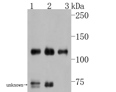
Fig1:; Western blot analysis of TAG1 on different lysates. Proteins were transferred to a PVDF membrane and blocked with 5% BSA in PBS for 1 hour at room temperature. The primary antibody ( 1/500) was used in 5% BSA at room temperature for 2 hours. Goat Anti-Rabbit IgG - HRP Secondary Antibody (HA1001) at 1:5,000 dilution was used for 1 hour at room temperature.; Positive control:; Lane 1: Mouse cerebellum tissue lysate; Lane 2: Human brain tissue lysate; Lane 3: Rat cerebellum tissue lysate
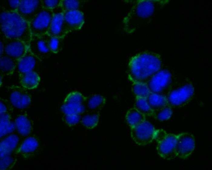
Fig2:; ICC staining of TAG1 in F9 cells (green). Formalin fixed cells were permeabilized with 0.1% Triton X-100 in TBS for 10 minutes at room temperature and blocked with 1% Blocker BSA for 15 minutes at room temperature. Cells were probed with the primary antibody ( 1/200) for 1 hour at room temperature, washed with PBS. Alexa Fluor®488 Goat anti-Rabbit IgG was used as the secondary antibody at 1/1,000 dilution. The nuclear counter stain is DAPI (blue).
- Background
-
References
- von Landenberg N. et. al. Conditional analyses of recurrence and progression in patients with TaG1 non-muscle-invasive bladder cancer. Urol Oncol. 2018 May
- Zhen YB. et. al. Expression of TAG1/APP signaling pathway in the proliferation and differentiation of glioma stem cells. Oncol Lett. 2017 Aug
Related Products / Services
Please note: All products are "FOR RESEARCH USE ONLY AND ARE NOT INTENDED FOR DIAGNOSTIC OR THERAPEUTIC USE"