
-
Product Name
Anti-CLIC2 antibody
- Documents
-
Description
Mouse monoclonal antibody to CLIC2
-
Tested applications
WB, ICC, IHC-P, FC
-
Species reactivity
Human
-
Alternative names
CLCNL2 antibody; CLIC2b antibody; MRXS32 antibody; XAP121 antibody
-
Isotype
Mouse IgG1
-
Preparation
This antigen of this antibody was recombinant protein within human clic2 aa 50-247 / 247.
-
Clonality
Monoclonal
-
Formulation
Liquid, 1*TBS (pH7.4), 0.2% BSA, 50% Glycerol. Preservative: 0.05% Sodium Azide.
-
Storage instructions
Store at +4℃ after thawing. Aliquot store at -20℃. Avoid repeated freeze / thaw cycles.
-
Applications
WB: 1:500-1:2,000
ICC: 1:50-1:100
IHC-P: 1:100-1:500
FC: 1:100-1:500
-
Validations
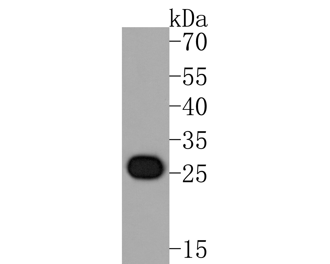
Fig1:; Western blot analysis of CLIC2 on K562 cell lysates. Proteins were transferred to a PVDF membrane and blocked with 5% NFDM/TBST for 1 hour at room temperature. The primary antibody ( 1/500) was used in 5% NFDM/TBST at room temperature for 2 hours. Goat Anti-Mouse IgG - HRP Secondary Antibody (HA1006) at 1:20,000 dilution was used for 1 hour at room temperature.
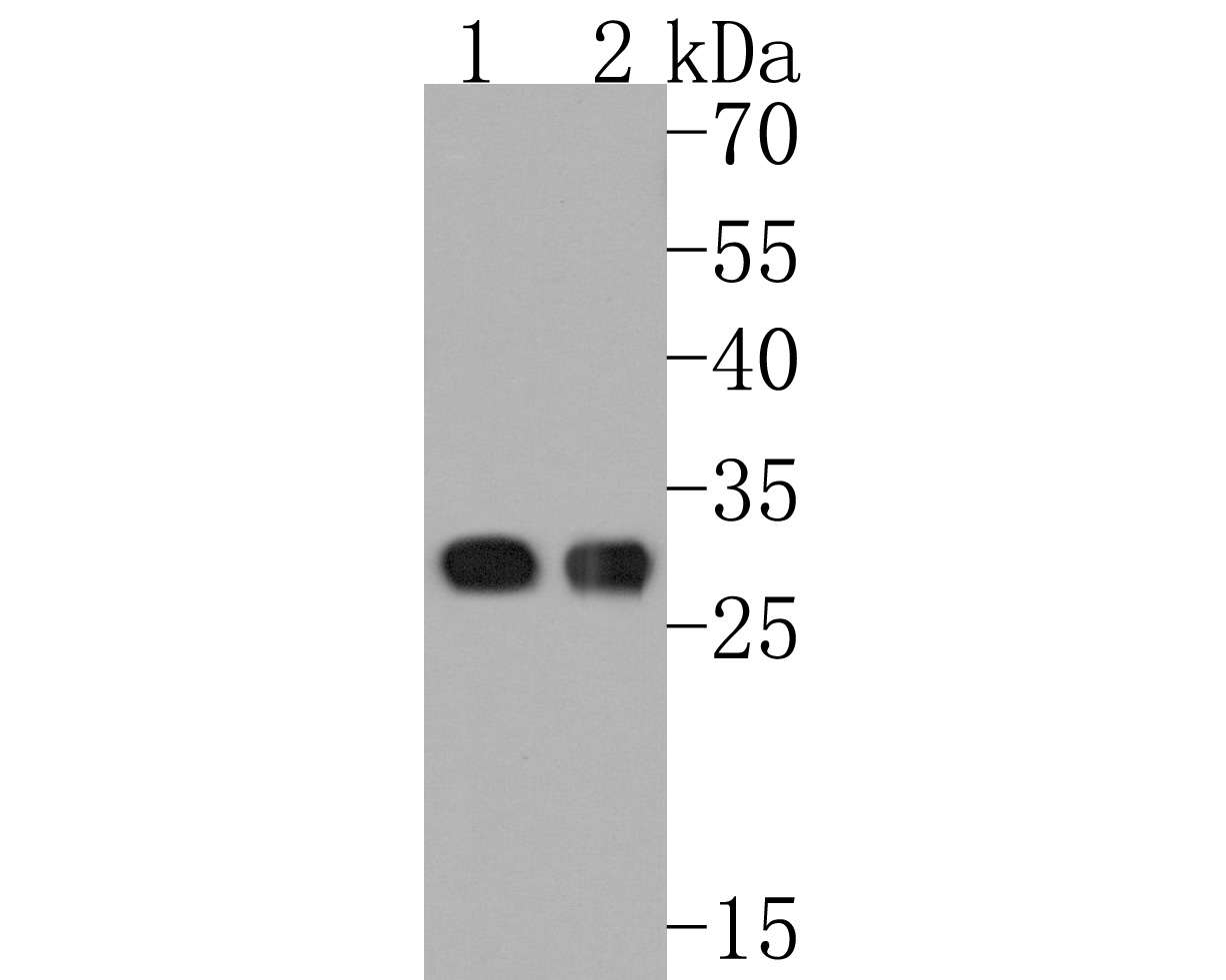
Fig2:; Western blot analysis of CLIC2 on different lysates. Proteins were transferred to a PVDF membrane and blocked with 5% NFDM/TBST for 1 hour at room temperature. The primary antibody ( 1/500) was used in 5% NFDM/TBST at room temperature for 2 hours. Goat Anti-Mouse IgG - HRP Secondary Antibody (HA1006) at 1:20,000 dilution was used for 1 hour at room temperature.; Positive control:; Lane 1: Human liver tissue lysate; Lane 2: Human skeletal muscle tissue lysate
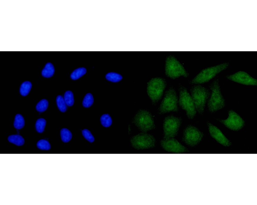
Fig3:; ICC staining of CLIC2 in HepG2 cells (green). Formalin fixed cells were permeabilized with 0.1% Triton X-100 in TBS for 10 minutes at room temperature and blocked with 10% negative goat serum for 15 minutes at room temperature. Cells were probed with the primary antibody ( 1/50) for 1 hour at room temperature, washed with PBS. Alexa Fluor®488 conjugate-Goat anti-Mouse IgG was used as the secondary antibody at 1/1,000 dilution. The nuclear counter stain is DAPI (blue).
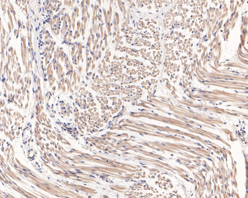
Fig4:; Immunohistochemical analysis of paraffin-embedded human fetal skeletal muscle tissue using anti-CLIC2 antibody. The section was pre-treated using heat mediated antigen retrieval with Tris-EDTA buffer (pH 9.0) for 20 minutes.The tissues were blocked in 1% BSA for 30 minutes at room temperature, washed with ddH; 2; O and PBS, and then probed with the primary antibody ( 1/400) for 30 minutes at room temperature. The detection was performed using an HRP conjugated compact polymer system. DAB was used as the chromogen. Tissues were counterstained with hematoxylin and mounted with DPX.
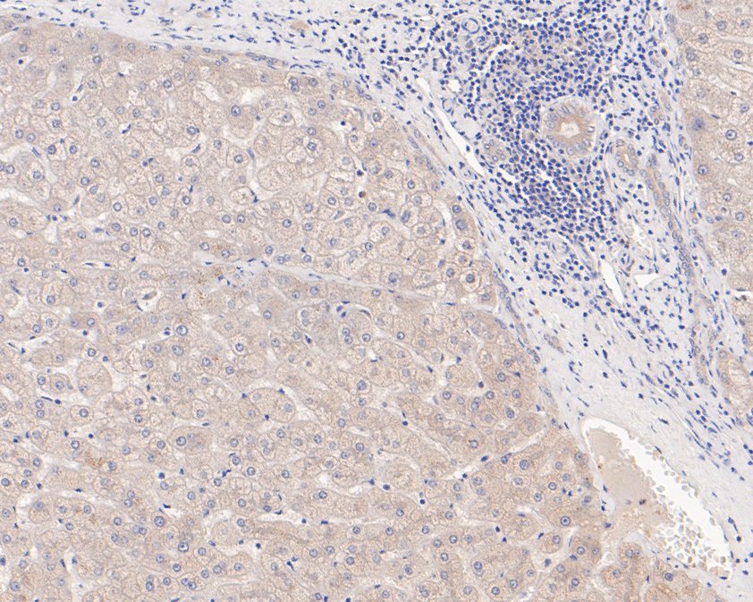
Fig5:; Immunohistochemical analysis of paraffin-embedded human liver tissue using anti-CLIC2 antibody. The section was pre-treated using heat mediated antigen retrieval with Tris-EDTA buffer (pH 9.0) for 20 minutes.The tissues were blocked in 1% BSA for 30 minutes at room temperature, washed with ddH; 2; O and PBS, and then probed with the primary antibody ( 1/400) for 30 minutes at room temperature. The detection was performed using an HRP conjugated compact polymer system. DAB was used as the chromogen. Tissues were counterstained with hematoxylin and mounted with DPX.
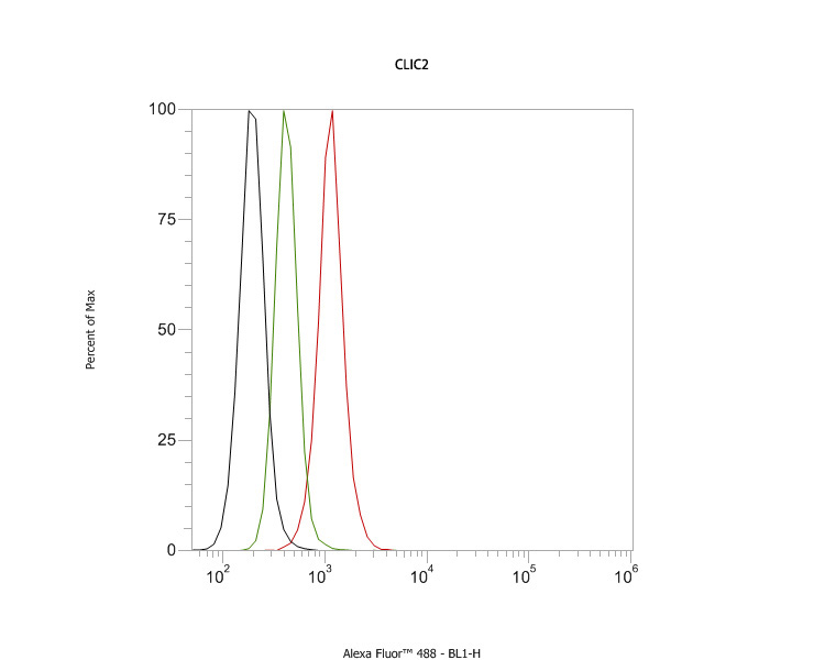
Fig6:; Flow cytometric analysis of CLIC2 was done on SH-SY5Y cells. The cells were fixed, permeabilized and stained with the primary antibody ( 1ug/ml) (red) compared with Mouse IgG, monoclonal - Isotype Control ( green). After incubation of the primary antibody at room temperature for an hour, the cells were stained with a Alexa Fluor®488 conjugate-Goat anti-Mouse IgG Secondary antibody at 1/1000 dilution for 30 minutes.Unlabelled sample was used as a control (cells without incubation with primary antibody; black).
- Background
-
References
- Zeng J. et. al. Tilapia and human CLIC2 structures are highly conserved. Biochem Biophys Res Commun. 2018 Jan
- Syahidatulamali CS. et. al. Association of anti-CLIC2 and anti-HMGB1 autoantibodies with higher disease activity in systemic lupus erythematosus patients. J Postgrad Med. 2017 Oct-Dec
Related Products / Services
Please note: All products are "FOR RESEARCH USE ONLY AND ARE NOT INTENDED FOR DIAGNOSTIC OR THERAPEUTIC USE"