
-
Product Name
Anti-Cldn1 antibody
- Documents
-
Description
Rabbit polyclonal antibody to Cldn1
-
Tested applications
WB, IHC-P, FC
-
Species reactivity
Human, Mouse, Rat
-
Alternative names
AI596271 antibody
-
Isotype
Rabbit IgG
-
Preparation
This antigen of this antibody was klh conjugated synthetic peptide derived from mouse claudin 1 121-211/211
-
Clonality
Polyclonal
-
Formulation
Liquid, 0.01M TBS(pH7.4) with 1% BSA, 0.03% Proclin300 and 50% Glycerol.
-
Storage instructions
Store at -20℃ for one year. Avoid repeated freeze/thaw cycles. The lyophilized antibody is stable at room temperature for at least one month and for greater than a year when kept at -20℃. When reconstituted in sterile pH 7.4 0.01M PBS or diluent of antibody the antibody is stable for at least two weeks at 2-4℃.
-
Applications
WB:1:500-2000
IHC-P:1:400-800
FC:1µg/Test
-
Validations
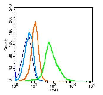
Fig1: Blank control: Raji(blue).; Primary Antibody:Rabbit Anti-Claudin 1 antibody , Dilution: 1μg in 100 μL 1X PBS containing 0.5% BSA;; Isotype Control Antibody: Rabbit IgG(orange) ,used under the same conditions );; Secondary Antibody: Goat anti-rabbit IgG-PE(white blue), Dilution: 1:200 in 1 X PBS containing 0.5% BSA.; Protocol; The cells were fixed with 2% paraformaldehyde (10 min). Antibody ( 1μg /1x10^6 cells) were incubated for 30 min on the ice, followed by 1 X PBS containing 0.5% BSA + 1 0% goat serum (15 min) to block non-specific protein-protein interactions. Then the Goat Anti-rabbit IgG/PE antibody was added into the blocking buffer mentioned above to react with the primary antibody of 175334# at 1/200 dilution for 30 min on ice. Acquisition of 20,000 events was performed.
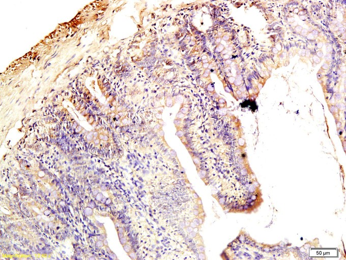
Fig2: Tissue/cell: rat intestine tissue; 4% Paraformaldehyde-fixed and paraffin-embedded;; Antigen retrieval: citrate buffer ( 0.01M, pH 6.0 ), Boiling bathing for 15min; Block endogenous peroxidase by 3% Hydrogen peroxide for 30min; Blocking buffer (normal goat serum,C-0005) at 37℃ for 20 min;; Incubation: Anti-Claudin-1 Polyclonal Antibody, Unconjugated 1:200, overnight at 4℃, followed by conjugation to the secondary antibody(SP-0023) and DAB(C-0010) staining
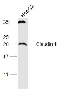
Fig3: Sample:; HepG2(Human) Cell Lysate at 30 ug; Primary: Anti-Claudin 1 at 1/1000 dilution; Secondary: IRDye800CW Goat Anti-Rabbit IgG at 1/20000 dilution; Predicted band size: 23 kD; Observed band size: 23 kD
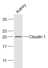
Fig4: Sample:; Kidney (Mouse) Lysate at 40 ug; Primary: Anti-Claudin 1 at 1/1000 dilution; Secondary: IRDye800CW Goat Anti-Rabbit IgG at 1/20000 dilution; Predicted band size: 23 kD; Observed band size: 23 kD
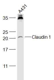
Fig5: Sample:; A431(Human) Cell Lysate at 30 ug; Primary: Anti-Claudin 1 at 1/1000 dilution; Secondary: IRDye800CW Goat Anti-Rabbit IgG at 1/20000 dilution; Predicted band size: 23 kD; Observed band size: 23 kD
- Background
Related Products / Services
Please note: All products are "FOR RESEARCH USE ONLY AND ARE NOT INTENDED FOR DIAGNOSTIC OR THERAPEUTIC USE"