
-
Product Name
Anti-CDK1 (3H7) Mouse antibody
- Documents
-
Description
CDK1 (3H7) Mouse monoclonal antibody
-
Tested applications
WB, ICC/IF, FC, IP
-
Species reactivity
Human
-
Isotype
Mouse IgG1
-
Preparation
Antigen: Purified recombinant fragment of CDC2 expressed in E. Coli.
-
Clonality
Monoclonal
-
Formulation
Ascitic fluid containing 0.03% sodium azide.
-
Storage instructions
Store at 4°C short term. Store at -20°C long term. Avoid freeze / thaw cycle.
-
Applications
WB: 1/500 - 1/2000
ICC: 1/200 - 1/1000
FC: 1/200 - 1/400
ELISA: 1/10000
-
Validations
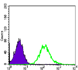
Flow cytometric analysis of PC-2 cells using CDC2 mouse mAb (green) and negative control (purple).
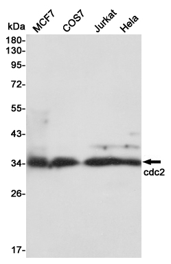
Western blot detection of cdc2 in MCF7,COS7,Jurkat and Hela,3T3 cell lysates using cdc2 mouse mAb (1:1000 diluted).Predicted band size:34KDa.Observed band size:34KDa.
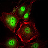
Immunofluorescence analysis of Hela cells using CDC2 mouse mAb (green). Red
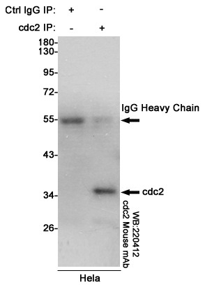
Immunoprecipitation analysis of Hela cell lysates using cdc2 mouse mAb.
-
Background
Swiss-Prot Acc.P06493.The cell division control protein cdc2, also known as cyclin-dependent kinase 1 (Cdk1) or p34/cdk1, plays a key role in the control of the eukaryotic cell cycle, where it is required for entry into S-phase and mitosis. Cdc2 exists as a complex with both cyclin A and cyclin B. The best characterized of these associations is the Cdc2 p34 cyclin B complex, which is required for the G2 to M phase transition. Activation of Cdc2 is controlled at several steps including cyclin binding and phosphorylation of threonine 161. However, the critical regulatory step in activating cdc2 during progression into mitosis appears to be dephosphorylation of Tyr15 and Tyr14. Phosphorylation at Tyr15 and inhibition of Cdc2 is carried out by WEE1 and MIK protein kinases while Tyr15 dephosphorylation and activation of Cdc2 is carried out by the cdc25 phosphatase. The isoform CDC2deltaT is found in breast cancer tissues. Furthermore, cdc2/Cdk1 is a key mediator of neuronal cell death in brain development and degeneration.
Related Products / Services
Please note: All products are "FOR RESEARCH USE ONLY AND ARE NOT INTENDED FOR DIAGNOSTIC OR THERAPEUTIC USE"