
-
Product Name
Anti-CD68 antibody
- Documents
-
Description
Mouse monoclonal antibody to CD68
-
Tested applications
WB, IHC-P, ICC, FC
-
Species reactivity
Human
-
Alternative names
GP110 antibody; LAMP4 antibody; SCARD1 antibody
-
Isotype
IgG1
-
Preparation
This antigen of this antibody was recombinant protein
-
Clonality
Monoclonal
-
Formulation
Liquid, 1*TBS (pH7.4), 1%BSA, 40%Glycerol. Preservative: 0.05% Sodium Azide.
-
Storage instructions
Store at +4℃ after thawing. Aliquot store at -20℃ or -80℃. Avoid repeated freeze / thaw cycles.
-
Applications
WB: 1:500-1:2,000
ICC: 1:200-1:1,000
IHC-P: 1:200-1:1,000
FC: 1:50-1:100
-
Validations
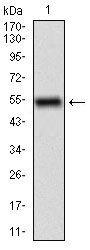
Fig1: Western blot analysis of CD68 on human CD68 recombinant protein using anti-CD68 antibody at 1/1,000 dilution.
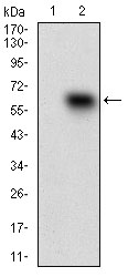
Fig2: Western blot analysis of CD68 on HEK293 (1) and CD68-hIgGFc transfected HEK293 (2) cell lysate using anti-CD68 antibody at 1/1,000 dilution.
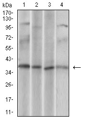
Fig3: Western blot analysis of CD68 on different cell lysate using anti-CD68 antibody at 1/1,000 dilution.; Positive control: Line1: U937 Line1: Hela Line2: HepG2 Line3: Jurkat
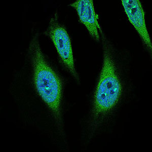
Fig4: ICC staining CD68 (green) and Actin filaments (red) in Hela cells. The nuclear counter stain is DAPI (blue). Cells were fixed in paraformaldehyde, permeabilised with 0.25% Triton X100/PBS.
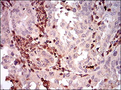
Fig5: Immunohistochemical analysis of paraffin-embedded human endometrial cancer tissue using anti-CD68 antibody. Counter stained with hematoxylin.
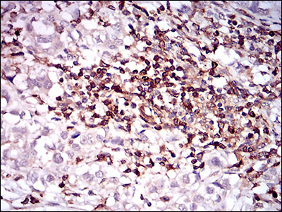
Fig6: Immunohistochemical analysis of paraffin-embedded human bladder cancer tissue using anti-CD68 antibody. Counter stained with hematoxylin.
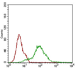
Fig7: Flow cytometric analysis of Hela cells with CD68 antibody at 1/100 dilution (green) compared with an unlabelled control (cells without incubation with primary antibody; red).
- Background
-
References
- Cao S et al. Hydrogen sulfide attenuates brain edema in early brain injury after subarachnoid hemorrhage in rats: Possible involvement of MMP-9 induced blood-brain barrier disruption and AQP4 expression. Neurosci Lett 621:88-97 (2016).
- Qi X et al. Development of inCVAX, In situ Cancer Vaccine, and Its Immune Response in Mice with Hepatocellular Cancer. J Clin Cell Immunol 7:N/A (2016).
Related Products / Services
Please note: All products are "FOR RESEARCH USE ONLY AND ARE NOT INTENDED FOR DIAGNOSTIC OR THERAPEUTIC USE"