
-
Product Name
Anti-Calreticulin (6H1) Mouse antibody
- Documents
-
Description
Calreticulin (6H1) Mouse monoclonal antibody
-
Tested applications
WB, IHC-P, ICC/IF
-
Species reactivity
Human, Mouse
-
Isotype
IgG2a
-
Preparation
Antigen: Synthetic peptide corresponding to aa (EEEDVPGQAKDELC) of human Calreticulin, conjugated to KLH.
-
Clonality
Monoclonal
-
Formulation
Ascitic fluid containing 0.03% sodium azide.
-
Storage instructions
Store at 4°C short term. Store at -20°C long term. Avoid freeze / thaw cycle.
-
Applications
WB: 1/500 - 1/2000
IHC: 1/200 - 1/1000
ICC: 1/200 - 1/1000
ELISA: 1/10000
-
Validations
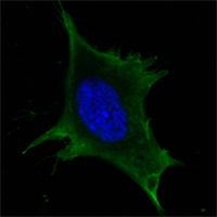
Confocal immunofluorescence analysis of 3T3-L1 cells using Calreticulin mouse mAb(green). Blue: DRAQ5 fluorescent DNA dye.
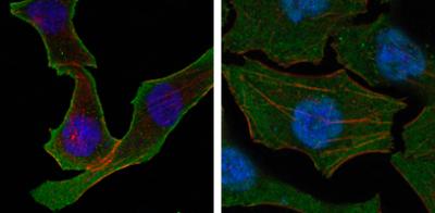
Confocal immunofluorescence analysis of SKBR-3 (left) and A549 (right) cells using Calreticulin mouse mAb (green). Red: Actin filaments have been labeled with DY-554 phalloidin. Blue: DRAQ5 fluorescent DNA dye.
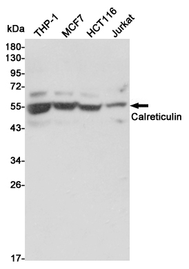
Western blot detection of Calreticulin in THP-1,MCF7,HCT116 and Jurkat cell lysates using Calreticulin mouse mAb (1:3000 diluted).Predicted band size:48KDa.Observed band size:55KDa.
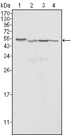
Western blot analysis using Calreticulin mouse mAb against Hela (1), A549 (2), NTERA2 (3) and MCF-7 (4) cell lysate.
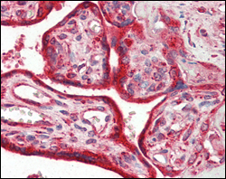
Immunohistochemical analysis of paraffin-embedded human placenta tissues using Calreticulin mouse mAb.
-
Background
Swiss-Prot Acc.P27797.Calreticulin, also known as RO, CRT, SSA, cC1qR, FLJ26680, CALR. Entrez Protein NP_004334. It is a multifunctional protein that acts as a major Ca(2+)-binding (storage) protein in the lumen of the endoplasmic reticulum. It is also found in the nucleus, suggesting that it may have a role in transcription regulation. Calreticulin binds to the synthetic peptide KLGFFKR, which is almost identical to an amino acid sequence in the DNA-binding domain of the superfamily of nuclear receptors. Calreticulin binds to antibodies in certain sera of systemic lupus and Sjogren patients which contain anti-Ro/SSA antibodies, it is highly conserved among species, and it is located in the endoplasmic and sarcoplasmic reticulum where it may bind calcium. The amino terminus of calreticulin interacts with the DNA-binding domain of the glucocorticoid receptor and prevents the receptor from binding to its specific glucocorticoid response element. Calreticulin can inhibit the binding of androgen receptor to its hormone-responsive DNA element and can inhibit androgen receptor and retinoic acid receptor transcriptional activities in vivo, as well as retinoic acid-induced neuronal differentiation. Thus, calreticulin can act as an important modulator of the regulation of gene transcription by nuclear hormone receptors. Systemic lupus erythematosus is associated with increased autoantibody titers against calreticulin but calreticulin is not a Ro/SS-A antigen. Earlier papers referred to calreticulin as an Ro/SS-A antigen but this was later disproven. Increased autoantibody titer against human calreticulin is found in infants with complete congenital heart block of both the IgG and IgM classes.
Related Products / Services
Please note: All products are "FOR RESEARCH USE ONLY AND ARE NOT INTENDED FOR DIAGNOSTIC OR THERAPEUTIC USE"