
-
Product Name
Anti-CACNA1C antibody
- Documents
-
Description
Rabbit polyclonal antibody to CACNA1C
-
Tested applications
Dot blot, ICC, IHC-P, FC
-
Species reactivity
Human, Mouse, Rat
-
Alternative names
TS antibody; LQT8 antibody; CACH2 antibody; CACN2 antibody; CaV1.2 antibody; CCHL1A1 antibody; CACNL1A1 antibody; TS. LQT8 antibody
-
Isotype
Rabbit IgG
-
Preparation
This antigen of this antibody was synthetic peptide within human cacna1c aa 800-880.
-
Clonality
Polyclonal
-
Formulation
Liquid, 1*PBS (pH7.4), 0.2% BSA, 50% Glycerol. Preservative: 0.05% Sodium Azide.
-
Storage instructions
Store at +4℃ after thawing. Aliquot store at -20℃. Avoid repeated freeze / thaw cycles.
-
Applications
Dot Blot: 1:500-1:1,000
ICC: 1:500-1:2,000
IHC-P: 1:50-1:200
FC: 1:50-1:100
-
Validations
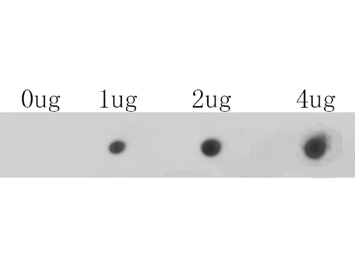
Fig1: Dot blot analysis of anti-CACNA1C on PVDF. 1ug, 2ug and 4ug of immunization peptides were given in this test. Anti-CACNA1C antibody was diluted with 1/500.
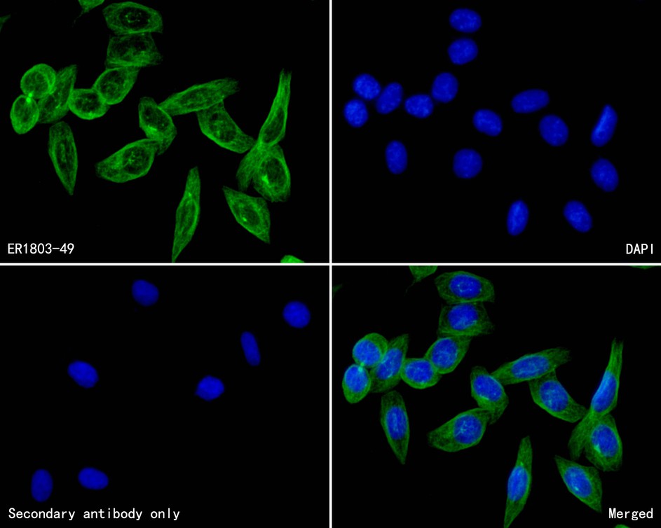
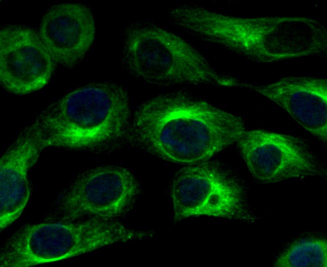
Fig2: ICC staining CACNA1C in SKOV-3 cells (green). The nuclear counter stain is DAPI (blue). Cells were fixed in paraformaldehyde, permeabilised with 0.25% Triton X100/PBS.
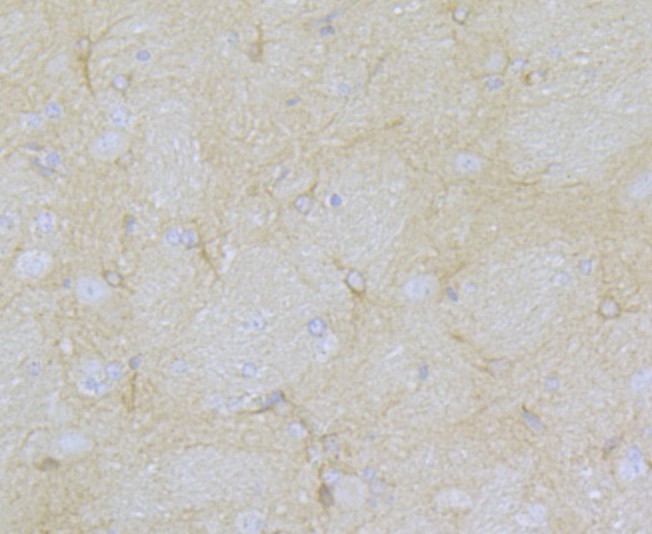
Fig3: Immunohistochemical analysis of paraffin-embedded rat brain tissue using anti-CACNA1C antibody. Counter stained with hematoxylin.
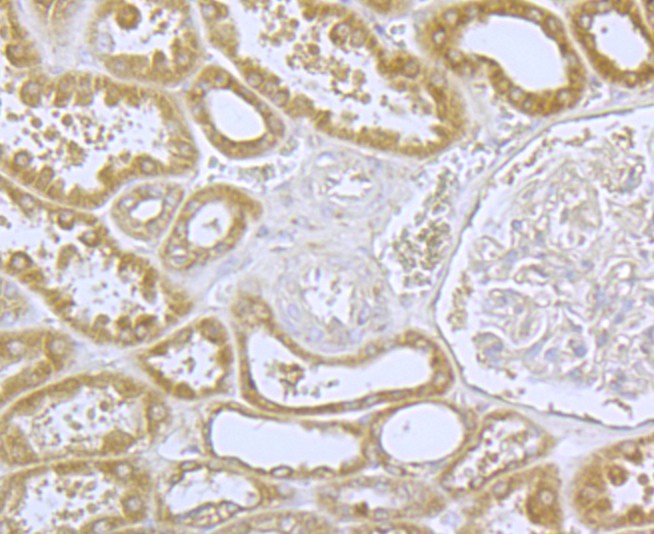
Fig4: Immunohistochemical analysis of paraffin-embedded human kidney tissue using anti-CACNA1C antibody. Counter stained with hematoxylin.
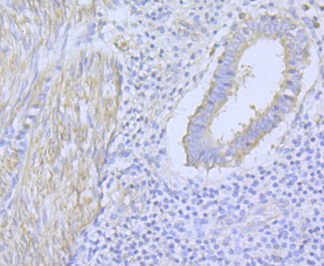
Fig5: Immunohistochemical analysis of paraffin-embedded human uterus tissue using anti-CACNA1C antibody. Counter stained with hematoxylin.
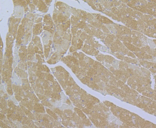
Fig6: Immunohistochemical analysis of paraffin-embedded mouse heart tissue using anti-CACNA1C antibody. Counter stained with hematoxylin.
Fig7: Flow cytometric analysis of SKOV-3 cells with CACNA1C antibody at 1/100 dilution (fuchsia) compared with an unlabelled control (cells without incubation with primary antibody; yellow). Alexa Fluor 488-conjugated goat anti-rabbit IgG was used as the
- Background
-
References
- Schultz D et al. Cloning, chromosomal localization, and functional expression of the alpha-1 subunit of the L-type voltage-dependent calcium channel from normal human heart. Proc Natl Acad Sci USA 90:6228-6232 (1993).
- Soldatov N M et al. Different voltage-dependent inhibition by dihydropyridines of human Ca2+ channel splice variants. J Biol Chem 270:10540-10543 (1995).
Related Products / Services
Please note: All products are "FOR RESEARCH USE ONLY AND ARE NOT INTENDED FOR DIAGNOSTIC OR THERAPEUTIC USE"