
-
Product Name
Anti-BCL2L12 antibody
- Documents
-
Description
Rabbit monoclonal antibody to BCL2L12
-
Tested applications
WB, ICC, IHC-P, FC
-
Species reactivity
Human
-
Isotype
Rabbit IgG
-
Preparation
This antigen of this antibody was recombinant protein within human bcl2l12 aa 150-300.
-
Clonality
Monoclonal
-
Formulation
Liquid, 1*TBS (pH7.4), 0.05% BSA, 40% Glycerol. Preservative: 0.05% Sodium Azide.
-
Storage instructions
Store at +4℃ after thawing. Aliquot store at -20℃. Avoid repeated freeze / thaw cycles.
-
Applications
WB:1:500-1:2,000
ICC:1:50-1:100
IHC-P:1:200-1:1000
FC:1:50-1:100
-
Validations
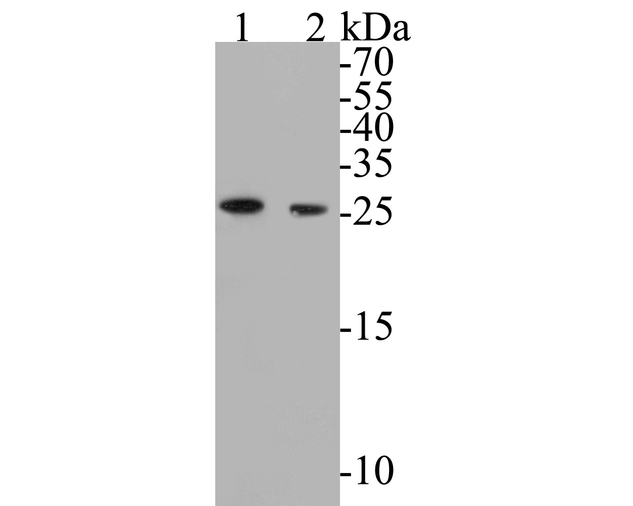
Fig1: Western blot analysis of BCL2L12 on different lysates. Proteins were transferred to a PVDF membrane and blocked with 5% BSA in PBS for 1 hour at room temperature. The primary antibody ( 1/500) was used in 5% BSA at room temperature for 2 hours. Goat Anti-Rabbit IgG - HRP Secondary Antibody (HA1001) at 1:5,000 dilution was used for 1 hour at room temperature.; Positive control:; Lane 1: MCF-7 cell lysates; Lane 2: PC-3M cell lysates
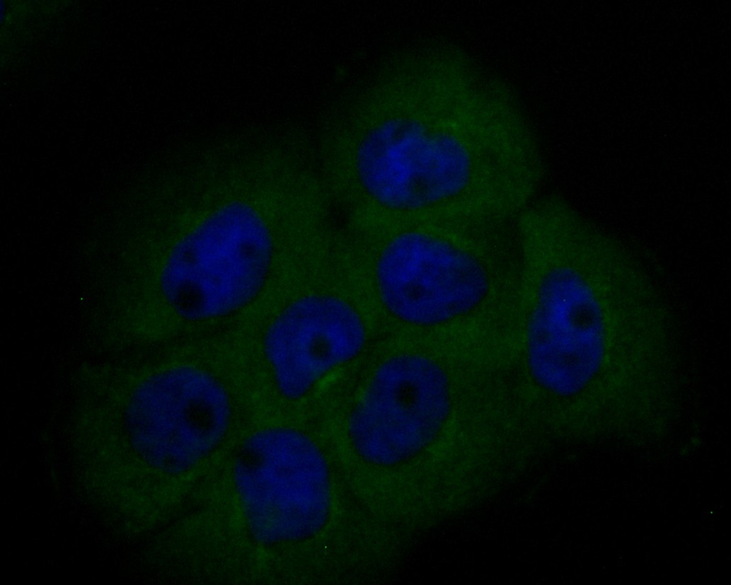
Fig2: ICC staining of BCL2L12 in A431 cells (green). Formalin fixed cells were permeabilized with 0.1% Triton X-100 in TBS for 10 minutes at room temperature and blocked with 1% Blocker BSA for 15 minutes at room temperature. Cells were probed with the primary antibody ( 1/100) for 1 hour at room temperature, washed with PBS. Alexa Fluor®488 Goat anti-Rabbit IgG was used as the secondary antibody at 1/1,000 dilution. The nuclear counter stain is DAPI (blue).
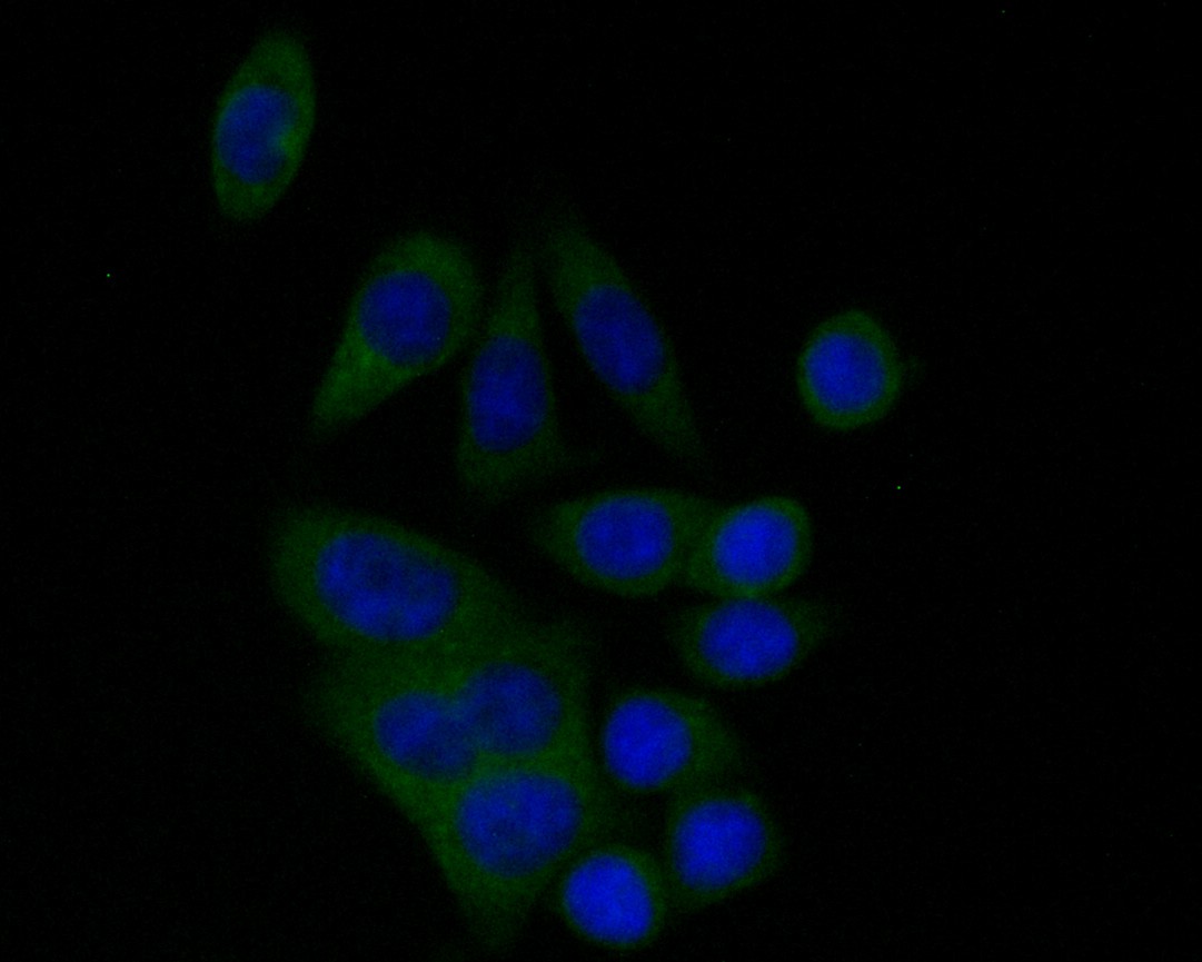
Fig3: ICC staining of BCL2L12 in SW620 cells (green). Formalin fixed cells were permeabilized with 0.1% Triton X-100 in TBS for 10 minutes at room temperature and blocked with 1% Blocker BSA for 15 minutes at room temperature. Cells were probed with the primary antibody ( 1/50) for 1 hour at room temperature, washed with PBS. Alexa Fluor®488 Goat anti-Rabbit IgG was used as the secondary antibody at 1/1,000 dilution. The nuclear counter stain is DAPI (blue).
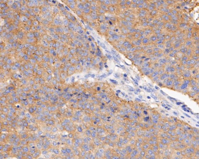
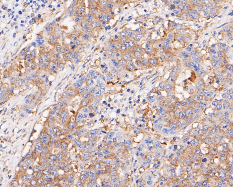
Fig4: Immunohistochemical analysis of paraffin-embedded human lung carcinoma tissue using anti-BCL2L12 antibody. The section was pre-treated using heat mediated antigen retrieval with sodium citrate buffer (pH 6.0) for 20 minutes. The tissues were blocked in 5% BSA for 30 minutes at room temperature, washed with ddH2O and PBS, and then probed with the primary antibody ( 1/400) for 30 minutes at room temperature. The detection was performed using an HRP conjugated compact polymer system. DAB was used as the chromogen. Tissues were counterstained with hematoxylin and mounted with DPX.
Fig5: Immunohistochemical analysis of paraffin-embedded human gastric carcinoma tissue using anti-BCL2L12 antibody. The section was pre-treated using heat mediated antigen retrieval with sodium citrate buffer (pH 6.0) for 20 minutes. The tissues were blocked in 5% BSA for 30 minutes at room temperature, washed with ddH2O and PBS, and then probed with the primary antibody ( 1/800) for 30 minutes at room temperature. The detection was performed using an HRP conjugated compact polymer system. DAB was used as the chromogen. Tissues were counterstained with hematoxylin and mounted with DPX.
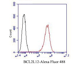
Fig6: Flow cytometric analysis of BCL2L12 was done on A431 cells. The cells were fixed, permeabilized and stained with the primary antibody ( 1/50) (red). After incubation of the primary antibody at room temperature for an hour, the cells were stained with a Alexa Fluor 488-conjugated Goat anti-Rabbit IgG Secondary antibody at 1/1000 dilution for 30 minutes.Unlabelled sample was used as a control (cells without incubation with primary antibody; black).
- Background
-
References
- Feng BS. et. al. Bcl2L12 mediates effects of protease-activated receptor-2 on the pathogenesis of Th2-dominated responses of patients with ulcerative colitis. Arch Biochem Biophys. 2018 Nov 1;657:8-14.
- Li MG. et. al. Bcl2L12 Contributes to Th2-Biased Inflammation in the Intestinal Mucosa by Regulating CD4+ T Cell Activities. J Immunol. 2018 Jul 15;201(2):725-733.
Related Products / Services
Please note: All products are "FOR RESEARCH USE ONLY AND ARE NOT INTENDED FOR DIAGNOSTIC OR THERAPEUTIC USE"