
-
Product Name
Anti-AGO3 antibody
- Documents
-
Description
Rabbit monoclonal antibody to AGO3
-
Tested applications
WB, ICC/IF, IHC-P, FC
-
Species reactivity
Human, Mouse, Rat
-
Alternative names
EIF2C3 antibody
-
Isotype
Rabbit IgG
-
Preparation
This antigen of this antibody was recombinant protein
-
Clonality
Monoclonal
-
Formulation
Liquid, 1*TBS (pH7.4), 0.05% BSA, 40% Glycerol. Preservative: 0.05% Sodium Azide.
-
Storage instructions
Store at +4℃ after thawing. Aliquot store at -20℃ or -80℃. Avoid repeated freeze / thaw cycles.
-
Applications
WB: 1:1,000
ICC/IF: 1:100-1:500
IHC-P: 1:50-1:200
FC: 1:50-1:100
-
Validations
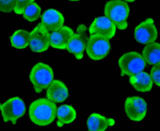
Fig1: ICC staining EIF2C3 in N2A cells (green). The nuclear counter stain is DAPI (blue). Cells were fixed in paraformaldehyde, permeabilised with 0.25% Triton X100/PBS.
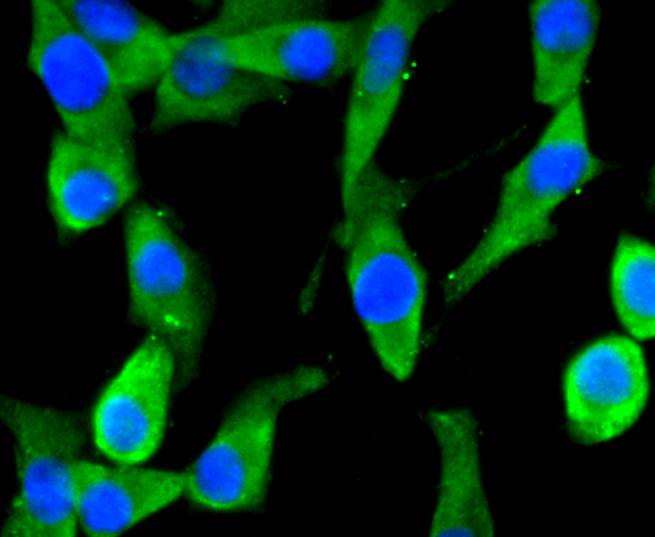
Fig2: ICC staining EIF2C3 in SHG-44 cells (green). The nuclear counter stain is DAPI (blue). Cells were fixed in paraformaldehyde, permeabilised with 0.25% Triton X100/PBS.
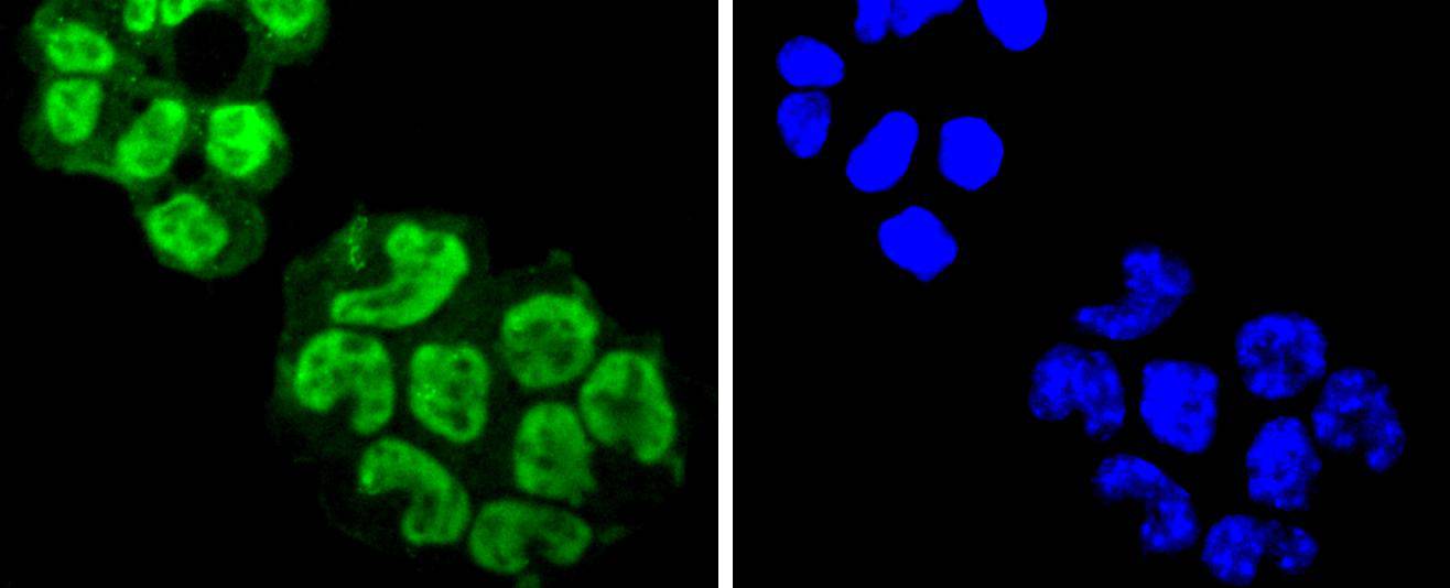
Fig3: ICC staining EIF2C3 in F9 cells (green). The nuclear counter stain is DAPI (blue). Cells were fixed in paraformaldehyde, permeabilised with 0.25% Triton X100/PBS.
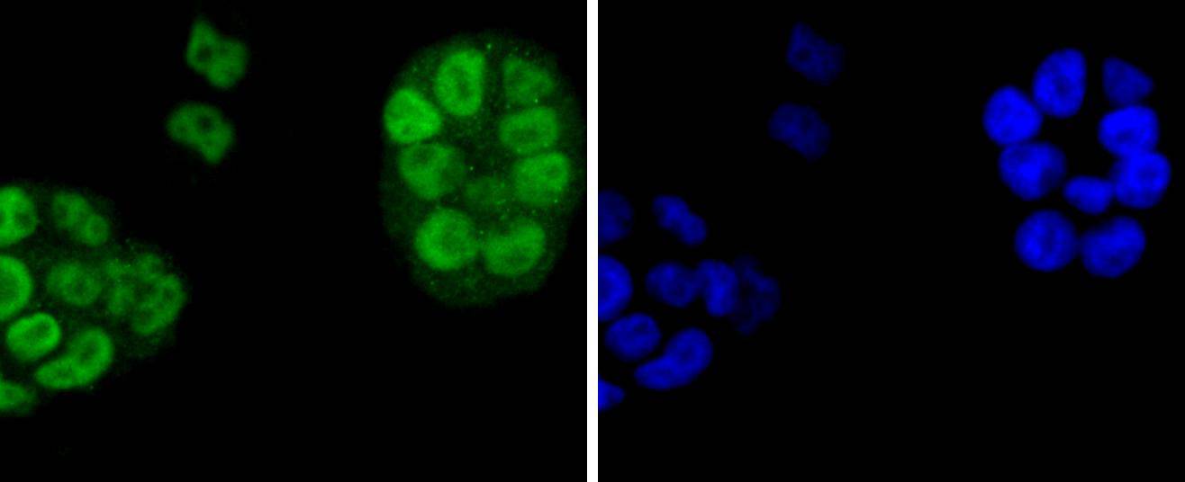
Fig4: ICC staining EIF2C3 in NCCIT cells (green). The nuclear counter stain is DAPI (blue). Cells were fixed in paraformaldehyde, permeabilised with 0.25% Triton X100/PBS.
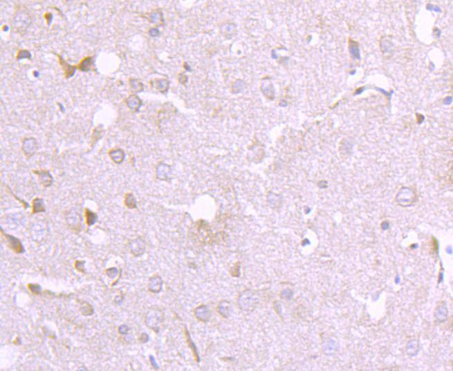
Fig5: Immunohistochemical analysis of paraffin-embedded rat spinal cord tissue using anti-EIF2C3 antibody. Counter stained with hematoxylin.
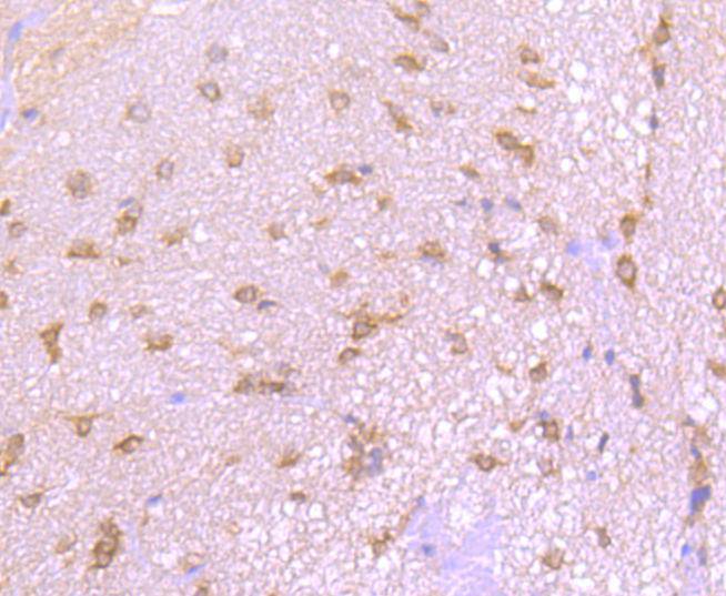
Fig6: Immunohistochemical analysis of paraffin-embedded rat brain tissue using anti-EIF2C3 antibody. Counter stained with hematoxylin.
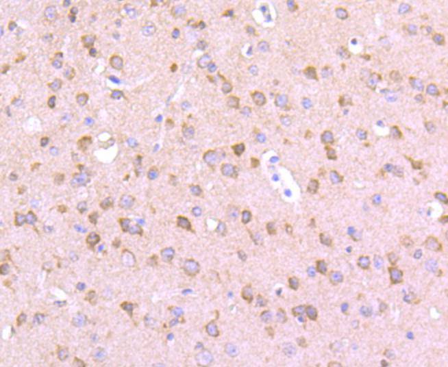
Fig7: Immunohistochemical analysis of paraffin-embedded mouse brain tissue using anti-EIF2C3 antibody. Counter stained with hematoxylin.
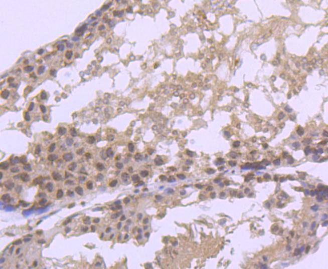
Fig8: Immunohistochemical analysis of paraffin-embedded rat testis tissue using anti-EIF2C3 antibody. Counter stained with hematoxylin.
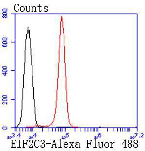
Fig9: Flow cytometric analysis of N2A cells with EIF2C3 antibody at 1/50 dilution (red) compared with an unlabelled control (cells without incubation with primary antibody; black). Alexa Fluor 488-conjugated goat anti rabbit IgG was used as the secondary
- Background
-
References
- Hein M.Y., et al. 2015. A human interactome in three quantitative dimensions organized by stoichiometries and abundances. Cell 163:712-723.
- Schurmann N., et al. 2013. Molecular dissection of human Argonaute proteins by DNA shuffling. Nat. Struct. Mol. Biol. 20:818-826.
Related Products / Services
Please note: All products are "FOR RESEARCH USE ONLY AND ARE NOT INTENDED FOR DIAGNOSTIC OR THERAPEUTIC USE"