
-
Product Name
LC3 antibody
- Documents
-
Description
LC3 Rabbit Polyclonal antibody. Positive FC detected in HeLa cells. Positive IHC detected in human gliomas tissue, human breast cancer tissue, human testis tissue. Positive IF detected in Starvation treated HEK-293 cells, Chloroquine treated HepG2 cells. Positive WB detected in Chloroquine treated HEK-293 cells, HEK-293 cells, HeLa cells, human skeletal muscle tissue, MCF7 cells, mouse brain tissue, mouse heart tissue, mouse liver tissue, mouse lung tissue, mouse testis tissue, NIH/3T3 cells, rat brain tissue. Positive IP detected in mouse brain tissue. Observed molecular weight by Western-blot: 16-18 kDa
-
Tested applications
ELISA, IHC, FC, IP, IF, WB
-
Species reactivity
Human, Mouse, Rat; other species not tested.
-
Alternative names
LC3 antibody; LC3B antibody; MAP1A/1BLC3 antibody; MAP1A/MAP1B LC3 B antibody; MAP1A/MAP1B light chain 3 B antibody; MAP1ALC3 antibody; MAP1LC3B antibody
-
Isotype
Rabbit IgG
-
Preparation
This antibody was obtained by immunization of LC3 recombinant protein (Accession Number: NM_022818). Purification method: Antigen affinity purified.
-
Clonality
Polyclonal
-
Formulation
PBS with 0.02% sodium azide and 50% glycerol pH 7.3.
-
Storage instructions
Store at -20℃. DO NOT ALIQUOT
-
Applications
Recommended Dilution:
WB: 1:500-1:5000
IP: 1:500-1:5000
IHC: 1:20-1:200
IF: 1:20-1:200
-
Validations
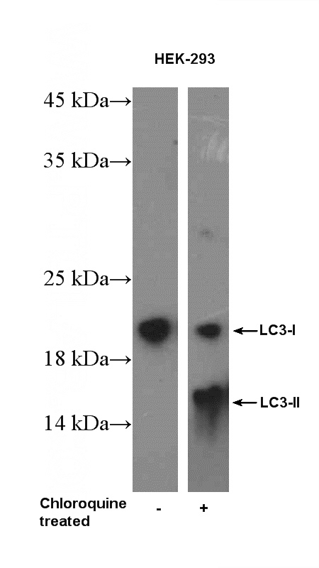
Chloroquine treated HEK-293 cells were subjected to SDS PAGE followed by western blot with Catalog No:112164(LC3 Antibody) at dilution of 1:1000
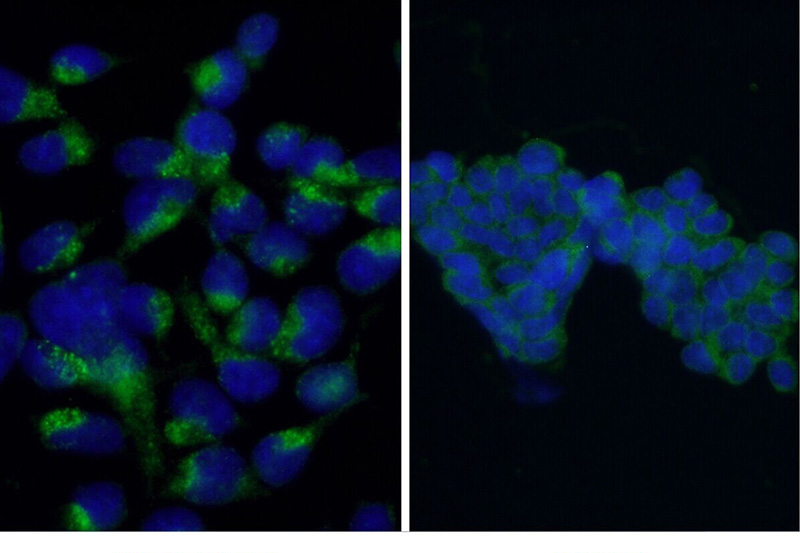
Immunofluorescent analysis of Starvation treated HEK-293 cells using Catalog No:112164 (LC3 Antibody) at dilution of 1:50 and Alexa Fluor 488-congugated AffiniPure Goat Anti-Rabbit IgG(H+L). 20 mM chloroquine was used to block the autophagy flux.
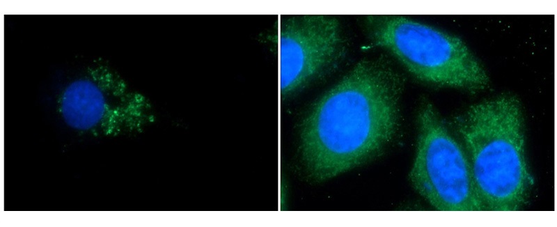
Immunofluorescent analysis of Chloroquine treated HepG2 cells and HepG2 cells using Catalog No:112164(LC3 Antibody) at dilution of 1:50 and Alexa Fluor 488-congugated AffiniPure Goat Anti-Rabbit IgG(H+L).
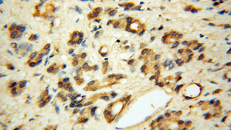
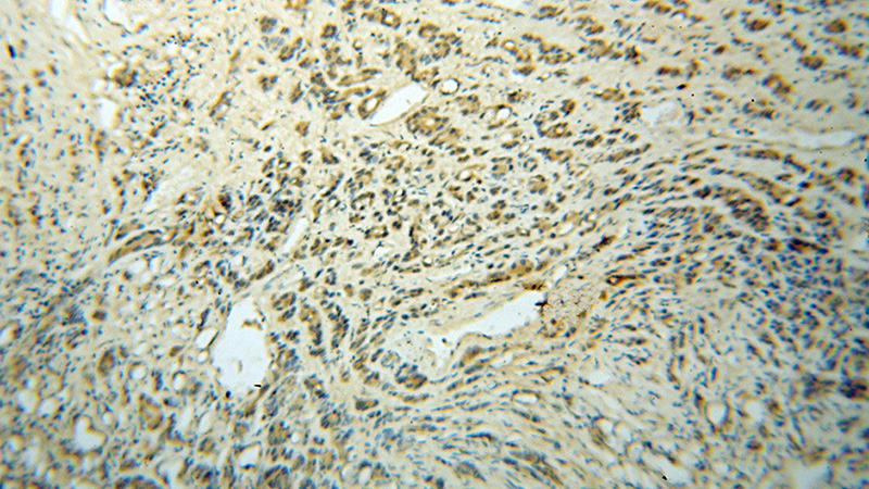
Immunohistochemical of paraffin-embedded human gliomas using Catalog No:112164 (LC3 antibody) at dilution of 1:50 (under 40x lens).
Immunohistochemical of paraffin-embedded human gliomas using Catalog No:112164(LC3 antibody) at dilution of 1:50 (under 10x lens)
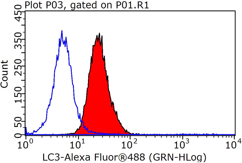
1X10^6 HeLa cells were stained with 0.2ug LC3 antibody (Catalog No:112164, red) and control antibody (blue). Fixed with 90% MeOH blocked with 3% BSA (30 min). Alexa Fluor 488-congugated AffiniPure Goat Anti-Rabbit IgG(H+L) with dilution 1:1000.
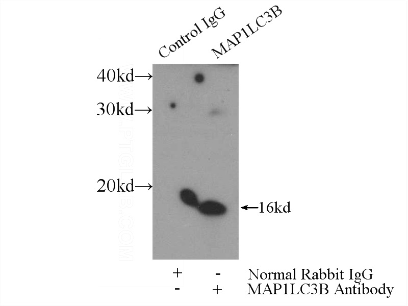
IP Result of anti-LC3 (IP:Catalog No:112164, 3ug; Detection:Catalog No:112164 1:1000) with mouse brain tissue lysate 4000ug.
-
Background
Map1LC3, also known as LC3, is the human homolog of yeast Apg8 and is involved in the formation of autophagosomal vacuoles, called autophagosomes. Three human Map1LC isoforms, MAP1LC3A, MAP1LC3B, and MAP1LC3C, undergo post-translational modifications during autophagy. And they differ in their post-translation modifications during autophagy. Map1LC3 also exists in two modified forms, an 18-kDa cytoplasmic form that was originally identified as a subunit of the microtubule-associated protein 1, and a 16-kDa form that is associated with the autophagosome membrane. This antibody can cross react with MAP1LC3A, MAP1LC3B, and MAP1LC3C.
-
References
- Wei DH, Jia XY, Liu YH. Cathepsin L stimulates autophagy and inhibits apoptosis of ox-LDL-induced endothelial cells: potential role in atherosclerosis. International journal of molecular medicine. 31(2):400-6. 2013.
- Deng X, Zhang F, Rui W. PM2.5-induced oxidative stress triggers autophagy in human lung epithelial A549 cells. Toxicology in vitro : an international journal published in association with BIBRA. 27(6):1762-70. 2013.
Related Products / Services
Please note: All products are "FOR RESEARCH USE ONLY AND ARE NOT INTENDED FOR DIAGNOSTIC OR THERAPEUTIC USE"