
-
Product Name
Anti-NF-kB p65 (3D2) Mouse antibody
- Documents
-
Description
NF-kB p65 (3D2) Mouse monoclonal antibody
-
Tested applications
WB, IHC-P, ICC/IF, IP
-
Species reactivity
Human, Mouse, Rat
-
Isotype
IgG1
-
Preparation
Antigen: Recombinant Protein of Transcription factor p65
-
Clonality
Monoclonal
-
Formulation
PBS, pH 7.4, containing 0.02% sodium azide as Preservative and 50% Glycerol.
-
Storage instructions
Store at 4°C short term. Store at -20°C long term. Avoid freeze / thaw cycle.
-
Applications
WB: 1/500-2000
IP:1/20
IF: 1/50-1/100
IHC: 1/50-300
-
Validations
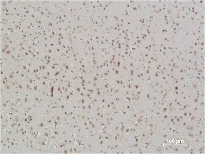
Immunohistochemical analysis of paraffin-embedded Rat Brain Tissue using NFkB p65 Mouse mAb diluted at 1:500.
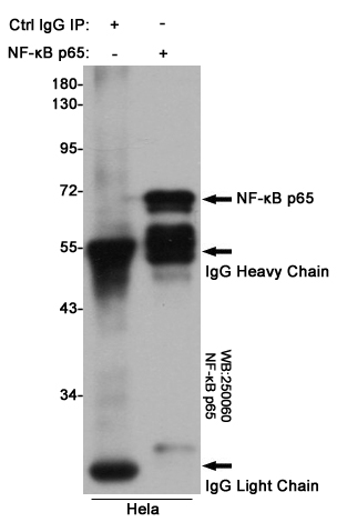
Immunoprecipitation analysis of Hela cell lysates using NF-κB p65 (3D2) Mouse mAb.
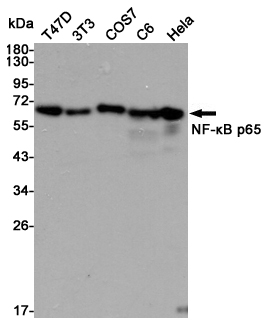
Western blot detection of NF-kB p65 in T47D,3T3,COS7,C6 and Hela cell lysates using NF-kB p65 mouse mAb (1:10000 diluted).Predicted band size:65kDa.Observed band size:65kDa.
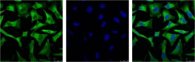
IF analysis of Hela and DAPI diluted at 1:100.
-
Background
Swiss-Prot Acc.Q04206.NF-kappa-B is a pleiotropic transcription factor present in almost all cell types and is the endpoint of a series of signal transduction events that are initiated by a vast array of stimuli related to many biological processes such as inflammation, immunity, differentiation, cell growth, tumorigenesis and apoptosis. NF-kappa-B is a homo- or heterodimeric complex formed by the Rel-like domain-containing proteins RELA/p65, RELB, NFKB1/p105, NFKB1/p50, REL and NFKB2/p52 and the heterodimeric p65-p50 complex appears to be most abundant one. The dimers bind at kappa-B sites in the DNA of their target genes and the individual dimers have distinct preferences for different kappa-B sites that they can bind with distinguishable affinity and specificity. Different dimer combinations act as transcriptional activators or repressors, respectively. NF-kappa-B is controlled by various mechanisms of post-translational modification and subcellular compartmentalization as well as by interactions with other cofactors or corepressors. NF-kappa-B complexes are held in the cytoplasm in an inactive state complexed with members of the NF-kappa-B inhibitor (I-kappa-B) family. In a conventional activation pathway, I-kappa-B is phosphorylated by I-kappa-B kinases (IKKs) in response to different activators, subsequently degraded thus liberating the active NF-kappa-B complex which translocates to the nucleus. NF-kappa-B heterodimeric p65-p50 and p65-c-Rel complexes are transcriptional activators. The NF-kappa-B p65-p65 complex appears to be involved in invasin-mediated activation of IL-8 expression. The inhibitory effect of I-kappa-B upon NF-kappa-B the cytoplasm is exerted primarily through the interaction with p65. p65 shows a weak DNA-binding site which could contribute directly to DNA binding in the NF-kappa-B complex. Associates with chromatin at the NF-kappa-B promoter region via association with DDX1. Essential for cytokine gene expression in T-cells (PubMed:15790681).
Related Products / Services
Please note: All products are "FOR RESEARCH USE ONLY AND ARE NOT INTENDED FOR DIAGNOSTIC OR THERAPEUTIC USE"