
-
Product Name
Anti-LPP (6F6) Mouse antibody
- Documents
-
Description
LPP (6F6) Mouse monoclonal antibody
-
Tested applications
WB, IHC-P, ICC/IF, IP
-
Species reactivity
Human, Mouse, Monkey, Hamster
-
Isotype
Mouse IgG1
-
Preparation
Antigen: Purified recombinant fragment of human LPP expressed in E. Coli.
-
Clonality
Monoclonal
-
Formulation
Ascitic fluid containing 0.03% sodium azide.
-
Storage instructions
Store at 4°C short term. Store at -20°C long term. Avoid freeze / thaw cycle.
-
Applications
WB: 1/500 - 1/2000
IHC: 1/200 - 1/1000
ICC: 1/200 - 1/1000
ELISA: 1/10000
-
Validations
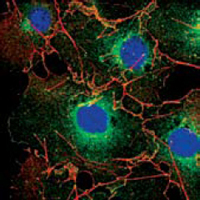
Confocal immunofluorescence analysis of COS cells using LPP mouse mAb (green). Red: Actin filaments have been labeled using DY-554 phalloidin. Blue: DRAQ5 fluorescent DNA dye.
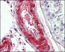
Immunohistochemical analysis of paraffin-embedded human vessels tissues using LPP mouse mAb.
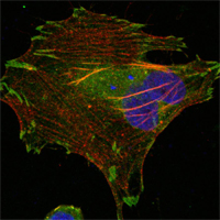
Confocal immunofluorescence analysis of Hela cells using LPP mouse mAb (green). Red: Actin filaments have been labeled using DY-554 phalloidin. Blue: DRAQ5 fluorescent DNA dye.
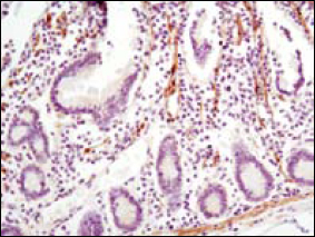
Immunohistochemical analysis of paraffin-embedded human small intestine using LPP mouse mAb with DAB staining.
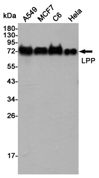
Western blot detection of LPP in A549,MCF7,C6 and Hela cell lysates using LPP mouse mAb (1:5000 diluted).Predicted band size:66KDa.Observed band size:75KDa.
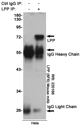
Immunoprecipitation analysis of Hela cell lysates using LPP (6F6) Mouse mAb.
-
Background
Swiss-Prot Acc.Q93052.LIM domain containing preferred translocation partner in lipoma. The Zyxin family of proteins contains five members, Ajuba, LIMD1, LPP,TRIP6 and Zyxin. LPP (LIM-containing lipoma-preferred partner), a LIM domain-containing scaffolding protein contains three LIM domains at its carboxyterminus, which are preceded by a proline-rich pre-LIM region containing a number of protein interaction domains. LPP, an 80 kDa protein, localizes to sites of cell adhesion, such as focal adhesions and cell-cell contacts,and shuttles to the nucleus where it has transcriptional activation capacity.The human LPP gene maps to chromosomal location 3q28, and preferentiallytranslocates to the HMGIC gene in a subclass of human benign mesenchymal tumors known as lipomas.
Related Products / Services
Please note: All products are "FOR RESEARCH USE ONLY AND ARE NOT INTENDED FOR DIAGNOSTIC OR THERAPEUTIC USE"