
-
Product Name
MAP2 antibody
- Documents
-
Description
MAP2 Rabbit Polyclonal antibody. Positive IF detected in Hela cells. Positive IHC detected in human brain tissue, mouse brain tissue, rat brain tissue. Positive IP detected in mouse brain tissue. Positive WB detected in SH-SY5Y cells, mouse brain tissue. Observed molecular weight by Western-blot: 280 kDa, 70-85 kDa
-
Tested applications
ELISA, WB, IF, IP, IHC
-
Species reactivity
Human,Mouse,Rat; other species not tested.
-
Alternative names
DKFZp686I2148 antibody; MAP 2 antibody; MAP2 antibody; MAP2A antibody; MAP2B antibody; MAP2C antibody
- Immunogen
-
Isotype
Rabbit IgG
-
Preparation
This antibody was obtained by immunization of MAP2 recombinant protein (Accession Number: XM_017004139). Purification method: Antigen affinity purified.
-
Clonality
Polyclonal
-
Formulation
PBS with 0.02% sodium azide and 50% glycerol pH 7.3.
-
Storage instructions
Store at -20℃. DO NOT ALIQUOT
-
Applications
Recommended Dilution:
WB: 1:500-1:5000
IP: 1:500-1:5000
IHC: 1:20-1:200
IF: 1:10-1:100
-
Validations
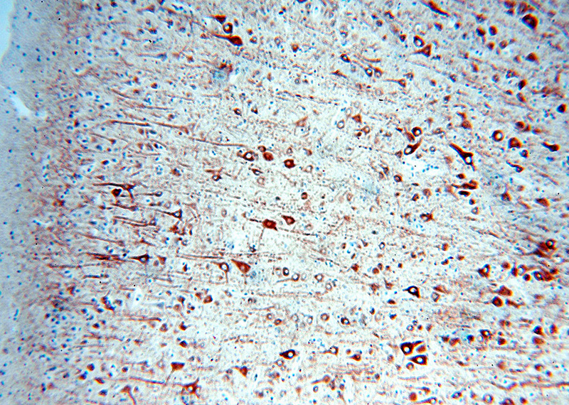
Immunohistochemical of paraffin-embedded human brain using Catalog No:112474(MAP2 antibody) at dilution of 1:100 (under 10x lens). Heat mediated antigen retrieved with Citric acid buffer, pH6.0.
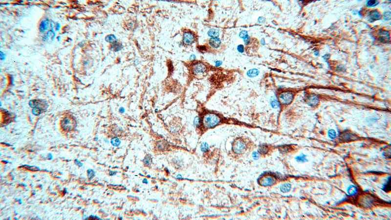
Immunohistochemical of paraffin-embedded human brain using Catalog No:112474(MAP2 antibody) at dilution of 1:100 (under 40x lens). Heat mediated antigen retrieved with Citric acid buffer, pH6.0.
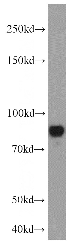
SH-SY5Y cells were subjected to SDS PAGE followed by western blot with Catalog No:112474(MAP2 antibody) at dilution of 1:1000
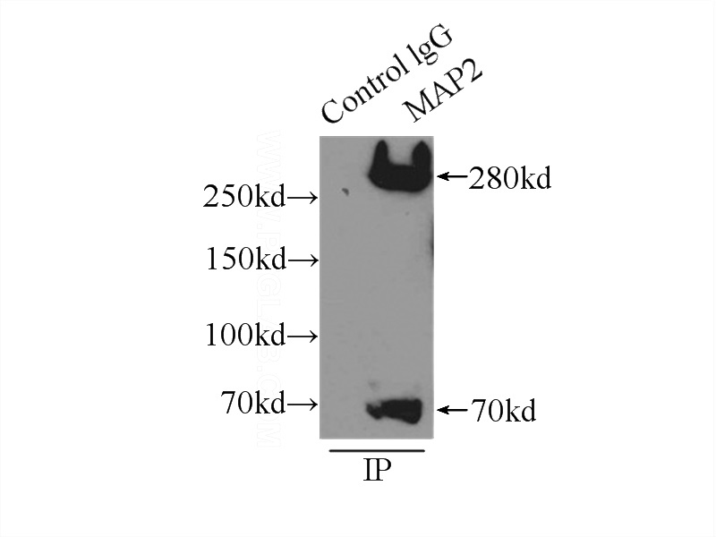
IP Result of anti-MAP2 (IP:Catalog No:112474, 3ug; Detection:Catalog No:112474 1:1000) with mouse brain tissue lysate 5000ug.
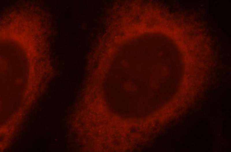
Immunofluorescent analysis of Hela cells, using Catalog No:112474 and Rhodamine-labeled goat anti-rabbit IgG (red).
-
Background
MAP2 (microtubule-associated protein 2) is a cytoskeleton protein abundant in brain and has important role in neuronal morphogenesis. Multiple high molecular weight (MW) and low molecular weight (MW) MAP2 isoforms are expressed within axons, dendrites, and cell bodies. The expression of MAP2 is regulated in both a tissue- and developmentally specific manner. The 280 kD MAP2B is present throughout rat brain development, and the slightly larger MAP2A appears first during the end of the second week of postnatal life. MAP2C, composed of several bands of about 70 kD, is present during early brain development, and largely disappears from the mature brain except for the retina, olfactory bulb, and cerebellum. MAP2 antibodies have been widely used to mark the neuron or dendrite formation. This antibody can recognize both high MW and low MW isoforms of MAP2.
-
References
- Gao J, Zhang C, Fu X. Effects of targeted suppression of glutaryl-CoA dehydrogenase by lentivirus-mediated shRNA and excessive intake of lysine on apoptosis in rat striatal neurons. PloS one. 8(5):e63084. 2013.
- Li Y, Yang M, Huang Z. AxonQuant: A Microfluidic Chamber Culture-Coupled Algorithm That Allows High-Throughput Quantification of Axonal Damage. Neuro-Signals. 22(1):14-29. 2014.
- Yin F, Guo L, Meng CY. Transplantation of mesenchymal stem cells exerts anti-apoptotic effects in adult rats after spinal cord ischemia-reperfusion injury. Brain research. 1561:1-10. 2014.
- Cicchetti F, Lacroix S, Cisbani G. Mutant huntingtin is present in neuronal grafts in Huntington disease patients. Annals of neurology. 76(1):31-42. 2014.
- Li Q, Dong C, Li W, Bu W, Wu J, Zhao W. Neuropeptide Y protects cerebral cortical neurons by regulating microglial immune function. Neural regeneration research. 9(9):959-67. 2014.
- Liu S, Li Z, Fu J. The effects of harvesting media on biological characteristics and repair potential of neural stem cells after traumatic brain injury. PloS one. 9(9):e107865. 2014.
- Yin F, Meng C, Lu R. Bone marrow mesenchymal stem cells repair spinal cord ischemia/reperfusion injury by promoting axonal growth and anti-autophagy. Neural regeneration research. 9(18):1665-71. 2014.
- Zhang L, Feng D, Tao H, DE X, Chang Q, Hu Q. Increased stathmin expression strengthens fear conditioning in epileptic rats. Biomedical reports. 3(1):28-32. 2015.
Related Products / Services
Please note: All products are "FOR RESEARCH USE ONLY AND ARE NOT INTENDED FOR DIAGNOSTIC OR THERAPEUTIC USE"