
-
Product Name
Anti-XRN2 antibody
- Documents
-
Description
Mouse monoclonal antibody to XRN2
-
Tested applications
WB, IHC-P
-
Species reactivity
Human
-
Isotype
Mouse IgG1
-
Preparation
This antigen of this antibody was recombinant protein
-
Clonality
Monoclonal
-
Formulation
Liquid, 1*TBS (pH7.4), 1%BSA, 40%Glycerol. Preservative: 0.05% Sodium Azide.
-
Storage instructions
Store at +4℃ after thawing. Aliquot store at -20℃ or -80℃. Avoid repeated freeze / thaw cycles.
-
Applications
WB: 1:500-1:2,000
IHC-P: 1:200-1:500
-
Validations
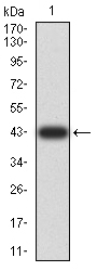
Fig1: Western blot analysis of XRN2 on human XRN2 recombinant protein using anti-XRN2 antibody at 1/1,000 dilution.
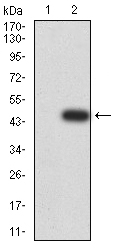
Fig2: Western blot analysis of XRN2 on HEK293 (1) and XRN2-hIgGFc transfected HEK293 (2) cell lysate using anti-XRN2 antibody at 1/1,000 dilution.
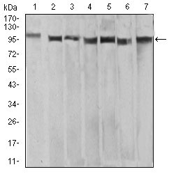
Fig3: Western blot analysis of XRN2 on different cell lysate using anti-XRN2 antibody at 1/1,000 dilution.; Positive control:; Lane 1: Raw264.7; Lane 2: HEK293; Lane 3: NTERA-2; Lane 4: LNcap; Lane 5: HepG2; Lane 6: HEK293; Lane 7: Hela
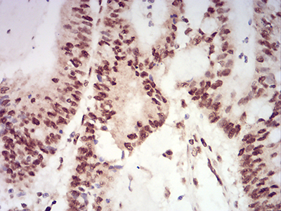
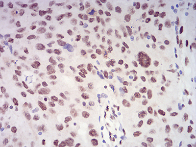
Fig4: Immunohistochemical analysis of paraffin-embedded human colon cancer tissue using anti-XRN2 antibody. Counter stained with hematoxylin.
Fig5: Immunohistochemical analysis of paraffin-embedded human ovarian cancer tissue using anti- XRN2 antibody. Counter stained with hematoxylin.
- Background
-
References
- Haas G et al. Identification of factors involved in target RNA-directed microRNA degradation. Nucleic Acids Res 44:2873-87 (2016).
- Gaulke CA et al. Intestinal Epithelial Barrier Disruption through Altered Mucosal MicroRNA Expression in Human Immunodeficiency Virus and Simian Immunodeficiency Virus Infections. J Virol 88:6268-80 (2014).
Related Products / Services
Please note: All products are "FOR RESEARCH USE ONLY AND ARE NOT INTENDED FOR DIAGNOSTIC OR THERAPEUTIC USE"